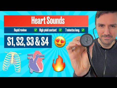
Heart sounds for beginners 🔥 🔥 🔥 S1, S2, S3 & S4 #heartsounds
1.61M views874 WordsCopy TextShare
The Learn Medicine Show
Heart sounds for beginners S1, S2, S3 & S4
*WATCH WITH THE BACKGROUND MUSIC REMOVED*;
https://youtu...
Video Transcript:
Hello, and welcome back. My name is dr coleman and in this tutorial, we're covering heart sounds so let's get straight down to the business. Heart sounds are usually presented in medical notes in an abbreviated form and they're presented as s1 s2 and s1 again the gap between s1 and s2 is known as systoli and this represents the period of time when the heart contracts the space between s2 and s1 is known as diastole and this represents the period of time when the heart relaxes this diagram is really useful because it depicts one movement through the
cardiac cycle and frames heart sounds temporally in relation to whether they occur during before or after heart contraction or relaxation let's take a closer look at the s1 and s2 heart sound the s1 heart sound is usually described as sounding like the word love and the s2 heart sound dub let's put the heart sounds in so you can appreciate this better let's now take a closer look at what causes the s1 and s2 heart sounds the s1 heart sound occurs when the tricuspid and mitral valves close simultaneously this is followed by ventricular contraction otherwise known
as systole and this forces blood through the pulmonary and aortic valves and the s2 heart sound occurs when the pulmonary and aortic valves close a period of heart muscle relaxation then occurs and this is known as diastole after which the cardiac cycle starts all over again let's now add in the heart sounds with this animation the four heart vowels and their respective heart sounds can be auscultated in the following anatomical areas let's now move on to s3 otherwise known as the third heart sound this heart sound occurs during early diastole just after s2 the word
kentucky is often used to illustrate the timing of the third heart sound in relation to s1 and s2 let's add in the heart sounds so you can appreciate this better to better understand how the s3 heart sound occurs we have to add in some blood flow to our animation the first heart sound is caused by the closure of the tricuspid and mitral valves and the second heart sound is caused by the closure of the pulmonary and aortic valves the s3 heart sound is produced by the rapid filling of the ventricles during diastole this produces audible
vibrations which are heard as the s3 heart sound let's now add the heart sounds to the animation we're going to start in slow motion but then we'll speed it up to real time [Music] the s3 heart sound is a low frequency sound heard best with the belt of the stethoscope at the apex of the heart it is heard in early diastole and is often referred to as a ventricular gallop it is caused by rapid ventricular filling which distends the ventricle and tortons papillary muscles the s3 heart sound is a physiological occurrence and can normally be
heard in children and in young healthy adults below the age of 30. but the s3 heart sound can also be an abnormal finding and typically can be found in pathology such as cardiomyopathies aortic and mitral regurgitation and constrictive pericarditis let's now take a look at the fourth heart sound otherwise known as s4 the s4 heart sound occurs just before s1 in late diastole the word tennessee is often used to help describe the rhythm produced by the added fourth heart sound let's add in the heart sound so you can better understand it let's now take a
closer look at why the fourth heart sound occurs now because the s4 heart sound occurs momentarily before s1 i have changed our graphical representation so that it now starts with diastole and ends with systole blood flows into the atria and ventricles during diastole and the fourth heart sound occurs when atrial contraction forces blood into abnormal non-compliant ventricles this produces audible vibrations which are heard as the s4 heart sound the s1 heart sound is produced by closure of the tricuspid and mitral valves and the s2 heart sound is produced by the closure of the pulmonary and
aortic valves let's add in the heart sounds now so that you can see this work we'll start this one in slow motion and then speed it up to real time [Music] the s4 heart sound is heard best at the cardiac apex with the bell of the stethoscope it is heard in late diastole and is often described as an atrial gallop rhythm it occurs due to blood being forced into non-compliant ventricles which means this heart sound only occurs in conditions where the ventricles are particularly stiff and do not relax normally the term diastolic dysfunction describes this
phenomenon so as a result the fourth heart sound is never physiological it can be heard abnormally in conditions such as hypertrophic cardiomyopathy and systemic hypertension now this brings us to the end of the tutorial if you found this video helpful make sure you smash that subscribe button and share this video with your friends as always thanks for stopping by i really appreciate it i will see you for the next tutorial
Related Videos

11:49
Heart murmurs for beginners 🔥 🔥 🔥 Part ...
The Learn Medicine Show
429,188 views

21:11
Heart Murmurs- in just 20 mins (use Headph...
Intellect Medicos
180,576 views
12:24
EKG/ECG Interpretation (Basic) : Easy and ...
MINT Nursing
6,388,370 views

5:13
Name that heart sound quiz ! Can You Ident...
The Learn Medicine Show
116,199 views

12:37
Chest X Rays (CXR) Made Easy! - Learn in 1...
Dr Ollie Burton
1,630,824 views

11:18
Lung Sounds- Normal & Abnormal (crackles, ...
Intellect Medicos
2,334,881 views

10:10
How To Suture: Intro To Suturing Like a Su...
Buck Parker, M.D.
4,036,859 views

1:55:11
Classical Music for Brain Power, Studying ...
Classical Mastermind
1,729,378 views

20:43
Abdominal organs (plastic anatomy)
Sam Webster
5,854,885 views
24:26
Types of IV Fluid - Fluid Management
ICU Advantage
1,955,679 views

11:10
How to Master the Cardiovascular Exam 🩺 |...
Lecturio Medical
559,802 views

8:43
The Cardiac Cycle is SO EASY! Stop Making ...
Interactive Biology
1,433,249 views

9:32
Respiratory Examination | OSCE Guide | UKM...
Geeky Medics
5,276,503 views
2:37:49
Mozart Effect in 432Hz – Boost Memory & Fo...
Classical Boost
550,262 views

16:15
HEART SOUNDS | S1, S2, S3, S4 (Use headpho...
Intellect Medicos
260,478 views

19:44
How to read a chest X-ray (in 20 mins) !
Intellect Medicos
2,804,045 views

40:13
#ecg interpretation : The animated Visual ...
The Learn Medicine Show
69,403 views

12:03
Lung Sounds Made Easy Nursing | Rhonchi, S...
RegisteredNurseRN
839,607 views

1:20:36
Evolution of Cardiac Diagnostics: A New Er...
Dr. Pradip Jamnadas, MD
946,723 views

16:33
Rapid, structured ECG interpretation: A vi...
The Learn Medicine Show
149,791 views