Structure of Microcirculation and Capillary System
7.27k views2118 WordsCopy TextShare
Nonstop Neuron
📝 All videos on Cardiovascular System: https://www.nonstopneuron.com/post/physiology-cardiovascular...
Video Transcript:
Welcome to nonstop neuron dot com, where learning medical concepts is as easy as watching cartoons. In this video, we will talk about the structure of the microcirculation and the capillary system. Let's get started.
Microcirculation, as per the name, is circulation at the micro level. It's basically the portion of the circulatory system from arterioles to capillaries to venules. Remember, it's this place where the exchange of nutrients and waste products which is the main function of the circulatory system happens.
So all the hard work of the heart and blood vessels is ultimately to serve this area. Now let's see these structures in more detail. The first thing to note is that the exact morphology varies from organ to organ depending on the needs or functions of that organ.
We will see the structure of an ideal microcirculation unit. It has an arteriole at one end, a venule at the other end, and a network of capillaries in between. Talking about the arteriole, they originate by branching of arteries.
Their wall contains a single continuous layer of smooth muscle cells. These smooth muscles receive innervation mainly by the sympathetic system. So the tone of arterioles is mainly under nervous control.
The capillaries originate from the arterioles. Right at its origin point, each capillary has a cuff of smooth muscle encircling the opening. It's called the precapillary sphincter.
It controls the entry of blood into the capillary. And it does not receive innervation. In some places, there are metarterioles connecting arterioles to venules.
They are somewhat larger than capillaries . Like arterioles, they also have smooth muscle fibers. But the layer is not continuous, and also they don't receive innervation.
Thus, both the metarterioles and precapillary sphincter do not receive innervation, and therefore are not controlled by the nervous system. However, they are very close to the cells they serve. So they respond directly to local conditions like oxygen concentration.
Isn't it amazing that the blood flow through even an individual capillary is also controlled? Now come to the capillary itself. Its internal diameter is 4 to 9 micrometers.
It's barely large enough for red blood cells and other cells to squeeze through. In fact, the small size of the capillary lumen brings the surface of RBCs close to the wall of the capillary. So the exchange of gases can happen rapidly.
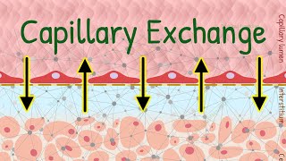
And that brings us to the wall of the capillary. In a cut section, the wall is like this. It is made up of a single layer of endothelial cells, and a basement membrane.
The endothelial cells are very thin except at the nucleus. To allow the exchange of material between the capillary and interstitium there are several pathways. One is the inter-cellular clefts.
This is space in between adjacent endothelial cells. This space contains proteins connecting the neighbor cells. But the fluid can freely pass through the gaps in between the proteins.
The cleft has a uniform width of about 6-7 nanometers. This size allows the filtration of most solutes ‒ except for large proteins like albumin. So this is one pathway for filtration.
The second pathway is not a physical pathway but rather a process. For this the endothelial cells have coated pits, that perform receptor-mediated endocytosis, capturing some fluid from the lumen. The formed vesicle then travels to the opposite side, and its content is released into the interstitium by exocytosis.
This is called trans-cytosis. Plasma proteins can enter the interstitium this way. Sometimes multiple vesicles fuse in series, and make a continuous passage through the cell.
This is called a vesicular channel, or trans-endothelial channel. And of course, the process of transcytosis and vesicular channels occur at multiple places in one endothelial cell. So this was about the passage through the endothelial cell.
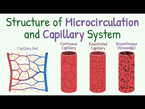
See, this is a general structure. Variations in this general morphology, can broadly create three different types of capillaries ‒with varying permeability. These types are continuous capillary, fenestrated capillary, and discontinuous capillary.
Continuous capillaries have an overall structure close to the general structure that we just studied. So comparatively we can say they have average permeability. Most organs in the body for example skeletal muscles, lungs, skin, etc have continuous capillaries.
In the brain, the extra tight continuous capillaries due to tight junctions between endothelial cells are less permeable. In fenestrated capillaries, the endothelial cells are thin, and have perforations called fenestrae. This increases the permeability of the capillary.
They are seen around the epithelia. For example the glomerular capillaries close to the epithelial cells of Bowman's capsule in the kidney are fenestrated. Finally, the sinusoidal or discontinuous capillaries are the most permeable.
In addition to fenestrae, they have large gaps between endothelial cells. They are found in sinusoids. For example, in bone marrow, the gaps between endothelial cells help newly formed blood cells enter the circulation.
So this was all about capillaries. On the other side, we have venules. Venules are larger in size than arterioles.
And they have weaker muscular coats. However, because the pressure in venules is low, it can contract to a considerable extent despite weak muscles. Venules converge to make veins, and then we have the vena cava.
So this is the structure of the capillary system. Now let's have a quick summary. Microcirculation as per the name, is made up of small blood vessels, starting from arteriole to venules.
It's this place where the exchange of substances between blood and tissue happens. The arteriolar smooth muscles are innervated. Metarterioles and precapillary sphincters respond to changes in local conditions.
The capillary wall is made up of a single layer of endothelial cells, and a basement membrane. To allow the passage of substances, there are gaps between the endothelial cells called intercellular clefts. Fluid can also cross the endothelial cell by transcytosis or through trans-endothelial channel.
Variation in this general structure is seen in different organs, depending on their specific needs. Broadly the types of capillaries are continuous, fenestrated, and sinusoidal, with increasing permeability in this order. That's all for the structure of the microcirculation and capillary system.
For watching the video until the end, I have some interesting facts for you. In the body, we have about ten billion of total capillaries. It's a huge number.
We literally have capillaries everywhere in the body. Hardly any functional cell is more than 30 micrometers away from a capillary. That's it for this video.
In this video, we will talk about the structure of the microcirculation and the capillary system. Let's get started. Microcirculation, as per the name, is circulation at the micro level.
It's basically the portion of the circulatory system from arterioles to capillaries to venules. Remember, it's this place where the exchange of nutrients and waste products which is the main function of the circulatory system happens. So all the hard work of the heart and blood vessels is ultimately to serve this area.
Now let's see these structures in more detail. The first thing to note is that the exact morphology varies from organ to organ depending on the needs or functions of that organ. We will see the structure of an ideal microcirculation unit.
It has an arteriole at one end, a venule at the other end, and a network of capillaries in between. Talking about the arteriole, they originate by branching of arteries. Their wall contains a single continuous layer of smooth muscle cells.
These smooth muscles receive innervation mainly by the sympathetic system. So the tone of arterioles is mainly under nervous control. The capillaries originate from the arterioles.
Right at its origin point, each capillary has a cuff of smooth muscle encircling the opening. It's called the precapillary sphincter. It controls the entry of blood into the capillary.
And it does not receive innervation. In some places, there are metarterioles connecting arterioles to venules. They are somewhat larger than capillaries .
Like arterioles, they also have smooth muscle fibers. But the layer is not continuous, and also they don't receive innervation. Thus, both the metarterioles and precapillary sphincter do not receive innervation, and therefore are not controlled by the nervous system.
However, they are very close to the cells they serve. So they respond directly to local conditions like oxygen concentration. Isn't it amazing that the blood flow through even an individual capillary is also controlled?
Now come to the capillary itself. Its internal diameter is 4 to 9 micrometers. It's barely large enough for red blood cells and other cells to squeeze through.
In fact, the small size of the capillary lumen brings the surface of RBCs close to the wall of the capillary. So the exchange of gases can happen rapidly. And that brings us to the wall of the capillary.
In a cut section, the wall is like this. It is made up of a single layer of endothelial cells, and a basement membrane. The endothelial cells are very thin except at the nucleus.
To allow the exchange of material between the capillary and interstitium there are several pathways. One is the inter-cellular clefts. This is space in between adjacent endothelial cells.
This space contains proteins connecting the neighbor cells. But the fluid can freely pass through the gaps in between the proteins. The cleft has a uniform width of about 6-7 nanometers.
This size allows the filtration of most solutes ‒ except for large proteins like albumin. So this is one pathway for filtration. The second pathway is not a physical pathway but rather a process.
For this the endothelial cells have coated pits, that perform receptor-mediated endocytosis, capturing some fluid from the lumen. The formed vesicle then travels to the opposite side, and its content is released into the interstitium by exocytosis. This is called trans-cytosis.
Plasma proteins can enter the interstitium this way. Sometimes multiple vesicles fuse in series, and make a continuous passage through the cell. This is called a vesicular channel, or trans-endothelial channel.
And of course, the process of transcytosis and vesicular channels occur at multiple places in one endothelial cell. So this was about the passage through the endothelial cell. See, this is a general structure.
Variations in this general morphology, can broadly create three different types of capillaries ‒with varying permeability. These types are continuous capillary, fenestrated capillary, and discontinuous capillary. Continuous capillaries have an overall structure close to the general structure that we just studied.
So comparatively we can say they have average permeability. Most organs in the body for example skeletal muscles, lungs, skin, etc have continuous capillaries. In the brain, the extra tight continuous capillaries due to tight junctions between endothelial cells are less permeable.
In fenestrated capillaries, the endothelial cells are thin, and have perforations called fenestrae. This increases the permeability of the capillary. They are seen around the epithelia.
For example the glomerular capillaries close to the epithelial cells of Bowman's capsule in the kidney are fenestrated. Finally, the sinusoidal or discontinuous capillaries are the most permeable. In addition to fenestrae, they have large gaps between endothelial cells.
They are found in sinusoids. For example, in bone marrow, the gaps between endothelial cells help newly formed blood cells enter the circulation. So this was all about capillaries.
On the other side, we have venules. Venules are larger in size than arterioles. And they have weaker muscular coats.
However, because the pressure in venules is low, it can contract to a considerable extent despite weak muscles. Venules converge to make veins, and then we have the vena cava. So this is the structure of the capillary system.
Now let's have a quick summary. Microcirculation as per the name, is made up of small blood vessels, starting from arteriole to venules. It's this place where the exchange of substances between blood and tissue happens.
The arteriolar smooth muscles are innervated. Metarterioles and precapillary sphincters respond to changes in local conditions. The capillary wall is made up of a single layer of endothelial cells, and a basement membrane.
To allow the passage of substances, there are gaps between the endothelial cells called intercellular clefts. Fluid can also cross the endothelial cell by transcytosis or through trans-endothelial channel. Variation in this general structure is seen in different organs, depending on their specific needs.
Broadly the types of capillaries are continuous, fenestrated, and sinusoidal, with increasing permeability in this order. That's all for the structure of the microcirculation and capillary system. For watching the video until the end, I have some interesting facts for you.
In the body, we have about ten billion of total capillaries. It's a huge number. We literally have capillaries everywhere in the body.
Hardly any functional cell is more than 30 micrometers away from a capillary. That's it for this video.
Related Videos
6:43
Capillary Exchange of Nutrients and Waste ...
Nonstop Neuron
6,049 views

20:32
Short Term Control of Local Blood Flow | C...
Nonstop Neuron
6,791 views

14:45
Capillary Exchange
Dr Matt & Dr Mike
49,982 views

15:10
Microcirculation in Capillaries
Dr. Najeeb Lectures
76,432 views
10:00
Blood Pressure, Blood Flow, Resistance and...
Nonstop Neuron
12,344 views
10:16
Vascular Compliance (Distensibility) & Its...
Nonstop Neuron
10,539 views
6:22
Structure of Blood Vessels | Layers of the...
Byte Size Med
54,397 views
17:31
Analysis of Cardiac Output Curve and Venou...
Nonstop Neuron
6,674 views

9:59
The Circulatory System Part 2: Blood Vessels
Professor Dave Explains
316,646 views

9:30
Blood Vessels, Part 1 - Form and Function:...
CrashCourse
2,877,256 views

7:13
Resistance to Blood Flow | Hemodynamics | ...
Nonstop Neuron
8,456 views

13:41
Arteries, Veins, and Blood Pressure
Siebert Science
138,262 views
22:38
Histology of the cardiovascular system
Human Anatomy Education
17,468 views
6:42
Why EXACTLY the Arteries are called Resist...
Nonstop Neuron
4,471 views

40:20
Cardiovascular | Fundamentals of Blood Pre...
Ninja Nerd
991,814 views
7:36
CARDIOVASCULAR REVIEW 3: CONTROL of BLOOD ...
Alila Medical Media
78,252 views

8:05
Types of blood vessels | Artery, capillary...
Learn Easy Science
218,668 views
44:32
Everything About Short-Term Regulation of ...
Nonstop Neuron
7,099 views

14:22
Starling Forces - made easy - Hydrostatic ...
Medicosis Perfectionalis
114,210 views

17:56
2024 ATI TEAS 7 Science Anatomy and Physio...
Nurse Cheung
6,594 views