Olfactory System: Anatomy and Physiology, Pathways, Animation.
438.35k views488 WordsCopy TextShare

Alila Medical Media
Physiology of Smell. Olfaction: A&P of special senses - sense of smell; loss of smell - anosmia; cli...
Video Transcript:
The olfactory system is responsible for the sense of smell, or olfaction. Basically, airborne molecules emitted by an odorant source are detected by olfactory sensory neurons located at the roof of the nasal cavity. These neurons convert chemical stimuli into electrical signals and send them via the olfactory nerve to the olfactory bulb, then to the brain, where they are interpreted as odors.
Odorant molecules are first dissolved in the mucus secreted by the olfactory epithelium, which guides them to the cilia of olfactory neurons. This is where odorant molecules bind to their receptors. Each neuron expresses a single type of protein receptor.
There are only about 400 different receptors in humans, but they are used in a combinatorial way such that one odorant can bind several receptors, and one receptor can bind several odorants. This enables the olfactory system to recognize an enormous number of odorants. Odorant receptors are G protein-coupled.
Upon binding to the odorant, a signaling cascade is activated, leading to membrane depolarization. When the olfactory stimulus is strong enough, action potentials are generated and conducted along the axon to the olfactory bulb. The axons of all olfactory sensory neurons form the olfactory nerve, also known as cranial nerve I.
In the olfactory bulb, these axons synapse with second-order neurons – the mitral and tufted cells, within structures called glomeruli. Each glomerulus receives axons from sensory neurons that express the same protein receptor. The second-order neurons are stimulated by sensory neurons, but they also receive inhibitory feedback from the cerebral cortex.
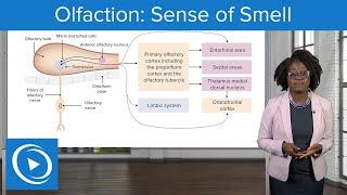
This means an odor can be perceived differently under different circumstances. For example, the smell of food is more appealing when one is hungry, and is less so when one is full. The axons of mitral and tufted cells form the olfactory tracts, which project directly to the primary olfactory cortex.
The primary olfactory cortex is not one but several cortical areas located on the base of the frontal lobe and inferior surface of the temporal lobe. These primary regions then project further to some other areas of the brain, mediating different aspects of odor recognition and response. Because olfactory neurons are exposed directly to the noxious external environment, they are replaced more often than other neurons.
Stem cells in the epithelium differentiate into new olfactory neurons, whose axons grow along the existing axons to the olfactory bulb. Any factors that destroy all olfactory neurons at once would result in permanent loss of sense of smell, a condition known as anosmia. Illnesses that cause inflammation of the nasal mucosa may lead to transient anosmia.
Loss of smell also affects the taste experience, as taste and smell are the 2 aspects of flavor. The ability to smell decreases with normal aging, but anosmia is also an early sign of several neurodegenerative disorders. Because epileptic seizures often originate from the brain area associated with the olfactory cortex, seizures are often preceded by hallucinations of disagreeable odors.
Related Videos
6:40
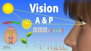
Vision: Anatomy and Physiology, Animation
Alila Medical Media
224,624 views
3:43
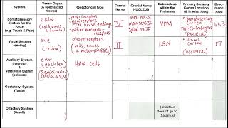
Sensory System Chart
M. Stabio
62 views

3:40
Non-Invasive Tech Helping Paralysis Patien...
WION
1,096 views
10:30
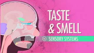
Taste & Smell: Crash Course Anatomy & Phys...
CrashCourse
2,518,297 views

4:20
How do we smell? - Rose Eveleth
TED-Ed
1,695,132 views
20:41
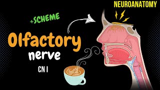
CN 1: Olfactory Nerve (Scheme, Pathway, Cl...
Taim Talks Med
60,424 views
8:25
Nature’s supercomputer lives on your dog |...
Big Think
431,003 views

10:43
Physiology Of Hearing Animation👂Understan...
Dr.G Bhanu Prakash Animated Medical Videos
101,157 views

24:00
Man Insults Keanu Reeves on a First Class ...
Heroic Acts
2,114,625 views

17:02
Dave Chappelle Stand-Up Monologue 2025 - SNL
Saturday Night Live
7,682,812 views

32:24
Neurology | Gustation (Taste Pathway)
Ninja Nerd
221,267 views

22:06
Olfactory system anatomy and physiology , ...
Physiology Open
6,607 views

12:46
Olfaction - structure and function | Proce...
khanacademymedicine
402,111 views
27:29
Neuroanatomy | Olfactory Nerve, Olfaction ...
Medicosis Perfectionalis
31,292 views

10:40
Hearing & Balance: Crash Course Anatomy & ...
CrashCourse
3,459,111 views
4:23
How Your Nose Works
Nemours KidsHealth
1,634,594 views
11:01
Nasal Anatomy (Cartilage, Nasal Cavity, Si...
Taim Talks Med
413,062 views

31:00
Neurology | Autonomic Nervous System
Ninja Nerd
1,929,027 views
6:18
Olfaction: Sense of Smell – Physiology | L...
Lecturio Nursing
20,062 views

37:51
Neurology | Olfactory Nerve: Cranial Nerve I
Ninja Nerd
464,003 views