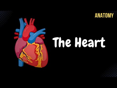
Anatomy of the Heart - External & Internal Structures
643.12k views3116 WordsCopy TextShare
Taim Talks Med
Content:
0:00 Introduction
0:33 Blood Circulation System
2:57 External Structures of the Heart
4:51 ...
Video Transcript:
Hey, What's up. Meditay here. Let's talk about the anatomy of the heart.
In this video, We're first going to look at how the blood circulates in the body. After that, we're going to cover the different external structures you'll find on the surface of the heart. Then we're going to open up the heart and go through the internal structures of each chamber, which include the right and left atrium and the right and left ventricles.
Then in the next video, we'll talk about the layers of the heart's wall, the conducting system, and then the topography. Now, let's start with the circulation. So here is see the anterior view of the thorax, right?
If you remove the sternum and the rib cartilage, you'll be able to see the heart right here. The heart lies between the pleura of the lungs in an area called the mediastinum. Now, let’s go ahead and pull the heart out.
So before we talk about the heart's anatomy, I wanna first take you through the general layout of the blood circulation in the body. Once you understand the concept of how the blood circulates in the body, you'll be able to learn the anatomy of the heart much more efficiently. Ok.
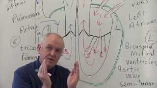
Throughout your body, your blood will either be deoxygenated, meaning it lacks oxygen. And Oxygenated blood that has oxygen. We’re going to start with the heart receiving deoxygenated blood from the body.
So the body has collected the oxygen, and it's sending the blood back towards the heart. The blood will enter the heart through the superior and inferior vena cava into the first chamber which is the right atrium. So the color blue will represent deoxygenated blood.
The blood is then going to go through the tricuspid valve into the right ventricle. The right ventricle is then going to pump the blood through the pulmonary valve and then into the pulmonary artery, which is then going to pump the blood to the lungs to exchange CO2 with Oxygen. And when this is done, blood will come back into the heart with oxygen, so the blood is oxygenated, now represented in red.
And they will come back through the pulmonary veins into the left atrium. The circulation where the blood goes from the heart to the lungs and back is called Pulmonary Circulation or the small circulation since it’s a circuit on its own. Now, the left atrium will send the blood through the bicuspid valve towards the left ventricle.
And when the left ventricle pump, it'll send the blood through the aortic valve where it's eventually going in end up in the aorta and then back to the body again. So the circulation in which the left ventricle pumps blood to the body, and the body sends the blood back into the heart is called the Systemic Circulation, or the major circulation. And notice that in the pulmonary circulation, Arteries contain blood without oxygen, and veins contain blood with oxygen.
Opposite from the systemic circulation, where arteries contain blood with oxygen and veins contain deoxygenated blood. Awesome. Let's finally start with the actual anatomy.
So if we turn this picture to the side, you’ll be able to see a pointy end called the Apex of the Heart, Which is directed forward and downwards to the left. And then if we turn the heart around and look at the backside. We can see the heart's base, or basis cordis, directed backward and a little to the right.
The base is connected with the great vessels you see here. So an apex and a base. Now the heart has three surfaces; it has Pulmonary Surface, towards the left lung, as you see here.
And then, we also have the Sternocostal Surface directed towards the inner surface of the ribs and the perfectly drawn sternum. And then, we have the Diaphragmatic Surface towards the central tendinous part of the diaphragm. Some sources say there are 5 surfaces on the heart.
Counting the right pulmonary surface and the base as two other surfaces, so sources may vary. Let's now take a closer look at the surface. The only margin that the heart has is the right margin or the right border, which lies between the Sternocostal Surface and the Diaphragmatic Surface.
One cool thing in knowing the external structures of the heart is that you’re actually able to know exactly where the inner chambers are based on the external structures. And I’ll show you how. First, we have the Coronary Sulcus, which separates the ventricles from the Atria.
This sulcus goes around, and here you see the posterior aspect of the heart again. The Coronary Sulcus goes all the way around like this separating the right and left atrium from the right and left ventricles. Another sulcus you’ll be able to see is the Anterior Interventricular Sulcus.
This sulcus continues posteriorly as the posterior interventricular sulcus, and it separates the right ventricle from the left ventricles externally. Now. Let’s take the heart and give it a good slice, then remove the upper half, and look at it from this perspective.
You’ll see that it looks like this. So we have this wall called the Septum of the Heart separating the right side of the heart from the heart's left side. And it's crucial to have this wall because remember the right side of the heart contains blood without oxygen, while the left side of the heart contains blood with oxygen, and you really don't mix those up, so it’s good to have this wall.
Alright, so we divide this wall into two parts. We first have the Interventricular Septum, separating the two ventricles. And we have the Interatrial Septum, separating the two atria.
Now the Interventricular septum can be further subdivided into two parts. We have the muscularis part down here since, in this area, the muscles are packed together. And up here is the membranous part, since this is the area where the valves are.
So that's mainly the general structures we have on the external heart. Now, let's look detailed into the different chambers. We'll start with the right atrium.
So here you see the anterior lateral view of the heart. And the right atrium is located right about here inside this area. In order to understand the full anatomy of the right atrium, we need to go through all six walls of it.
So let’s work our way through this list, starting with the Anterior Wall. So the Anterior Wall is right here, and it consists of the Right Auricle. The Right Auricle is a small muscle pouch, and this is where the Anterior and Lateral Wall has something in common.
Both have this muscle layer called Pectinate Muscle. As you see here, the pectinate muscle consists of striated muscle fibers, which help contract the right atrium to push the blood further into the right ventricle. That's the anterior and lateral walls so let's check them.
Now, let’s go ahead and remove the lateral and the anterior wall to look inside the right atrium. Here we can see a little bit of pectinate muscle we mentioned earlier. Now, if you look at the superior wall, the superior wall has an opening for the Superior Vena cava.
So That's mainly the superior wall. On the posterior wall, we can find the opening for the inferior vena cava. Remember, the heart is tilted forward, and because it’s tilted forward, the inferior vena cava becomes part of the posterior wall.
Another thing you’ll find in the posterior wall is, You know, anteriorly, you have the rough pectinati muscle, right? But posteriorly, between the opening for the superior and inferior vena cava, we have a wall called the Sinus of Vena Cava or the sinus venarum. This had a function during embryonic development, but now it forms the area for the SA node, for the conductive system.
Now, there's a line that separates the pectinate muscle and the sinus venarum. If we look at the outside of the lateral wall again, we have a crest called the terminal crest that separates the transition between the pectinate muscle and the sinus venarum. So that's mainly how we can distinguish where the sinus venarum is and where the pectinate muscle ends.
On the posterior wall, we can also see a valve. And this valve is called the valve for the inferior vena cava. It’s considered an embryonic valve that, during fetal life, the blood went directly from the inferior vena cava to a hole on the medial wall, which is now called the Oval Fossa.
The blood went directly from the right atrium to the left atrium since the fetus doesn’t need pulmonary circulation because they get their oxygen through the placenta. But in adults, the valve remains, it’s still there, but it remains as small folds on the posterior medial wall, which is that ridge you see here. So that is the posterior wall.
Now let’s do the medial wall. Remember, the medial wall is made up of the septum between the right and left atria. Called the Interatrial Septum.
On this septum, that’s where you'll find the oval fossa. And Around the oval fossa, you’ll see that it’s bordered by an elevation called border of oval fossa, also referred to as the limbus of oval fossa. Another thing you’ll find here is an opening for the coronary sinus, which lies between the medial and posterior wall.
This is the opening of the main vein draining blood from the heart. And this opening is guarded by a valve called The Valve for the coronary sinus. Which is really just a fold covering it.
So on the medial wall of the heart, you will also find very small openings returning blood directly from the layers of the heart's wall called the openings of the smallest cardiac veins. So that's mainly the structures on the medial wall. Now lastly, we have an inferior wall.
And to see this one, we need to slightly change the angle, as you see here. Now you’re able to see an opening called the right atrioventricular orifice or opening. That's the opening between the right atrium and the right ventricle.
And this opening is guarded by a tricuspid valve, which prevents the backflow of the blood. It's called a tricuspid valve because it has three cusps or three leaflets. So if we zoom in on it, you'll see three cusps right here—just a quick way to remember that the tricuspid valve lies on the right side.
The name tricuspid valve has the letters "R" "I," which resembles the word right, so the tricuspid valve is on the right side. And it consists of the anterior, posterior, and the septal cusps So that is the inferior wall. Now, let's look at the right ventricle.
So the right ventricle has a triangle shape, as you see here. Now on the base of the right ventricle, you'll find the Right Atrioventricular Opening, which is the opening between the right atrium and ventricle. Another opening you'll find is the Opening for the Pulmonary Trunk.
Now, If you look within the actual ventricle, you'll see a rough inner layer, right? And most of that rough inner layer is called Trabeculae carneae. The Trabeculae are essential because they prevent the blood from swirling around cuz remember, when the blood enters the heart, it enters with high velocity.
And by having this rough inner layer, you actually prevent the blood from getting air bubbles as they reach a sudden break which could cause a potential air embolism And here, you can also find papillary muscles, just like in the atria. Their function is connected to the tricuspid valve. So this valve again guards the right atrioventricular opening and has three cusps or leaflets, as I mentioned earlier.
It has the anterior cusp right here, and it has a posterior cusp and has a septal cusp towards the ventricular septum. That's a septum that divides the right and left ventricle. These cusps, including all other cusps, are formed by the endocardium.
We’ll talk more about that in the next video but keep that in mind. The opening is also surrounded by a fibrous ring that supports the valve and functions as an attachment point for these three cusps. So each cusp has 3 margins.
One margin of each cusp is attached to the fibrous ring. And the other two margins are attached to the Tendinous Chords, which are cords of fibers that attach the cusps to the papillary muscles, which control the valves' opening and closure. And then up here, we have something called the pulmonary valve, which has 3 semilunar cusps.
And these, just like the tricuspid valve, are formed by a double layer of the endocardium. If you’re unsure what endocardium is, that's no problem. For now, just know that the endocardium form these cusps.
The pulmonary valve also has a fibrous ring around where the base of the semilunar cusps are attached to. While the free margins are directed upwards into the pulmonary artery, as you see here. The pulmonary cusps have a free margin, as you see here, so they differ from the tricuspid valve.
There’s a nodule in the center of the free borders called Nodules of the semilunar valve, which provides a tight closure of the valves. And so here we can see another structure Between the semilunar valves and the walls of the pulmonary artery. We can see the sinuses called the sinus of the pulmonary trunk.
The pulmonary trunk is just another name for the pulmonary artery, and this sinus is basically just a space here that the cusps form. Here's another picture just to visualize it. You can see the 3 semilunar cusp down here, and you have a nodule in the middle which helps with the tight closure, and the space or the depression behind it's called the pulmonary sinus.
So that was the anatomy of the right side of the heart. Here again, you get the blood coming from the body into the right atrium. From the right atrium, blood goes through the tricuspid valve, as you see here, into the right ventricle.
Then the blood continues up through the pulmonary valve into the pulmonary artery. Which goes to the lungs, where it gets oxygenated, and then the oxygenated blood goes into the left atrium. So now, let's have a look at the walls of the left atrium.
Remember, the left atrium lies on the other side of the right atrium. Let’s repeat something first. So here you see an anterior view of the heart, where you’ll be able to see the right atrium again.
Remember, the anterior wall of the right atrium consists of the right auricle. But if you turn the heart so that we're looking at the posterior lateral surface. We can see that the anterior wall here is made up of the left auricle, which remember is a muscle pouch containing pectinate muscle.
And back here, we can find 4 blood vessels on the posterior wall where oxygenated blood from the lungs comes in. These blood vessels are called Pulmonary veins. So the posterior wall of the left atrium has four openings, called the Openings of the Pulmonary Veins.
Alright, so let's open up the walls and look at the inside. You can see that the medial wall is the Interatrial Septum, which, as you know, separates both Atria. And then, anteriorly, you'll find the opening, which leads into the left ventricle, The Left Atrioventricular Opening.
And this opening is also guarded by a valve called the bicuspid valve, or you can also use the term mitral valve. So that was all for the left atrium, now. Let's look at the left ventricle.
Here you see the left ventricle with the mitral valve. And if we tilt it a little bit in this direction, you will see one more valve. So let's now talk about the base of the left ventricle.
From these two angles, we're able to see the Left Atrioventricular Opening, where blood from the left atrium comes in, and the Aortic Opening, which leads into the aorta. Let's now do the Left Atrioventricular opening first and then do the Aortic Opening. Here you see the Left Atrioventricular Opening again, and it's guarded by the bicuspid valve or the mitral valve.
And you know, just like the other valves, it has a fibrous ring around that the cusps are attached to. Now since it’s called Bicuspid valve, it means that this valve consists of two leaflets. One is called the Anterior Cusp, and one is called Posterior Cusp.
But regardless of the name, there're actually several small cusps located in between them. So here we have the bicuspid valve. Here's the anterior cusp and here the posterior cusp.
And these small cusps right here, these are called commissural cusps, Which are very small cusps that support the valve. Now If you look closely at the surface of the left ventricle, you'll notice that it more or less has the same structures as the right ventricle. There's the Trabecula Carneae for the blood flow.
And here we have Papillary Muscles, which remember are attached to the Tendinous Chords. So the Tendinous Chords bind the cusps of the mitral valve onto the papillary muscles. So when the papillary muscles contract, they open up the mitral valve.
Now, let’s turn the heart in this direction so that we can see the aortic opening. This region that leads into the aortic opening is called the Aortic Vestibule inside the left ventricle. So this opening is guarded by the Aortic Valva, which is made up of three semilunar cusps, very similar to the pulmonary valve.
Here we’ll find three semilunar cusps, but this time, they’re called the Right semilunar cusp, the posterior semilunar cusp, and the Left semilunar cusp. So right posterior left. They also have nodules that help close the valve tightly.
We also have 3 aortic sinuses here on the top, which remember are just a depression formed by the semilunar valve. Awesome. So now we have gone through the pulmonary circulation, which pumps oxygenated blood into the left atrium.
The left atrium will now pump the blood through the bicuspid valve into the left ventricle, which will pump blood through the aortic valve into the Aorta. And then to the rest of the body. I really hope you found this video helpful.
If it did, please like, comment, share. Now in the next video, we'll look at the layers of the heart wall, the conducting system, and then the topography of the heart.
Related Videos
11:21
Anatomy of the Heart (Layers, Conducting S...
Taim Talks Med
202,092 views

23:14
Anatomy of the heart
Osmosis from Elsevier
87,337 views

26:26
Overview of Heart Anatomy Tutorial
The Noted Anatomist
392,804 views
13:06
Liver Anatomy (Function, Topography, Exter...
Taim Talks Med
597,747 views

13:46
NASA's SpaceX Crew-10 Docks With the ISS: ...
CNET
168,176 views

18:54
Can You Fool A Self Driving Car?
Mark Rober
6,806,742 views
29:14
Male Genital System (Internal & External) ...
Taim Talks Med
1,203,622 views

18:27
Forget Doping, These GENIUS Cheaters Will ...
RIFIANBOY
1,345,020 views

15:20
Heart Anatomy: Chambers, Valves, & Structu...
RegisteredNurseRN
56,940 views

22:27
I Rode the Fastest, Weirdest and Most Futu...
Ryan Trahan
2,316,417 views

3:17:34
Narendra Modi: Prime Minister of India - P...
Lex Fridman
152,250 views

21:55
Cardiovascular | Structures and Layers of ...
Ninja Nerd
1,189,042 views

9:35
Elon gets NIGHTMARE news about Tesla
Brian Tyler Cohen
1,161,008 views
21:33
Cardiovascular System 1, Heart, Structure ...
Dr. John Campbell
6,426,051 views
4:16
Gross anatomy of Right atrium (RA) Animati...
Dr.G Bhanu Prakash Animated Medical Videos
218,658 views

13:02
Race Highlights | 2025 Australian Grand Prix
FORMULA 1
4,279,898 views

29:41
I Found Axolotls in a Hidden Tunnel!
Bass fishing Productions
725,281 views

13:11
Trump gets UNEXPECTED SURPRISE in Greenland
Brian Tyler Cohen
908,669 views

5:07:37
A Warrior’s Rest – Peaceful Medieval Music...
Echoes Of Camelot
7,565 views

15:29
Coronary circulation of the heart
The Noted Anatomist
1,678,253 views