Neoplasia : Benign vs Malignant Tumors, Hallmarks, Spread & Clinical Manifestations of Cancer
190.62k views3916 WordsCopy TextShare

Med Today
Neoplasia, is an extremely important topic in general pathology.
Meaning of the word neoplasia is s...
Video Transcript:
in neoplasia neo means new and plagia means growth so simply the name neoplasia means new growth although all physicians know what they mean when they use the word neoplasia it has been difficult to develop an accurate definition before the development of molecular biology british oncologist willis made a close definition a neoplasm is an abnormal mass of tissue growth of which exceeds and is uncoordinated with normal tissues in the body and persists in the same excessive manner when the stimulus evoking the change is removed hope you can understand this definition the current definition implies that a
neoplasm is a disorder of cell growth that is triggered by a series of acquired mutations affecting a single cell and its clonal progeny let's try to understand what this means in the next slide any neoplasm in the body arises from a single cell that has undergone a genetic mutation therefore neoplasms are clonal in nature meaning that all the cells in a neoplasm consist of the same genetic composition these mutations give the neoplastic cells a survival and growth advantage resulting in abnormal excessive proliferation and producing an abnormal mass of tissue called a neoplasm proliferation of these
abnormal cells is independent of physiological growth stimuli of the body this is called the autonomous nature of neoplasms however neoplasms are dependent on the body for oxygen and nutrients tumor was the term originally applied to the swelling caused by inflammation however nowadays it is considered to be a synonym for the word neoplasm and the study of tumors is known as oncology could be either benign or malignant a tumor is said to be benign when its gross and microscopic appearances are relatively innocent implying that the tumor will remain localized and not spread to other sites benign
tumors are easily manageable with surgical removal so the patient generally survives however in some situations benign tumors may cause significant mobility and sometimes even death malignant tumors are collectively referred to as cancers they can invade and destroy adjacent structures and spread to distant sites which is known as metastasis malignant tumors are extremely fatal over benign tumors causing significant morbidity and mortality i will discuss about these in more detail in the following sections both benign and malignant tumors are composed of two essential components tumor parenchyma which consists of proliferating monoclonal neoplastic cells and reactive stroma which
consists of connective tissue blood vessels and cells of innate and adaptive immune system especially macrophages and lymphocytes the classification and behavior of tumors is based on the parenchymal component alternatively growth and spread of the tumor is critically dependent on the stromal tissue component in some tumors stromal support is scant thus the tumor looks soft and fleshy and in some tumors stromal support is high with abundant collagen this is called desmaplasia some tumors of the breast show desmaplasia now we know some basic facts about tumors and next let's discuss about the nomenclature of tumors let's take
benign tumors first most of the time the nine tumors are designated by attaching the suffix oma to the name of the cell type from which the tumor originates tumors of mesenchymal origin generally follow this rule for example benign tumor of fibrous tissue is called fibroma benign tumor of cartilage tissue is called chondroma benign tumor of smooth muscle tissue is called leo myoma and benign tumor of skeletal muscle tissue is called rhabdomyoma nomenclature of benign epithelial neoplasms is somewhat complex than others they are classified in several ways maybe according to their cells of origin or maybe
according to their microscopic pattern and architecture benign epithelial neoplasms derived from glandular epithelium are called adenomas one of the most common example is pituitary adenoma the nine epithelial neoplasms producing microscopically or macroscopically visible finger-like projections from epithelial surfaces are called papillomas and the nine epithelial neoplasms that produce cystic masses are called cystenomas commonest example is cystenoma of the ovary benign epithelial neoplasms producing papillary patterns that protrude into a cystic space are called papillary cyst adenomas commonly seen in the parroted gland there is another term in oncology called a polyp a polyp could be either a
benign or malignant tumor it is a macroscopically visible projection over a mucosal surface polyps are commonly seen in the colon and uterine endometrium the nomenclature of malignant tumors is not much different from benign tumors malignant tumors arising in mesenchymal tissues are called sarcomas for example malignant tumors of fibrous tissue are called fibrosarcomas malignant tumors of cartilage tissue are called chondrosarcomas malignant tumors of smooth muscle tissue are called leo myosarcomas and malignant tumors of skeletal muscle tissue are called rhabdomyosarcomas malignant tumors arising from blood forming cells are called leukemias or lymphomas and malignant tumors of epithelial
cell origin are called carcinomas for example squamous cell carcinoma is the malignant tumor arising in stratified squamous epithelium adenocarcinoma is the malignant tumor arising in glandular epithelium in addition there are also some tumors are named after benign sounding terms like lymphoma which is a malignant tumor but sounds like a benign one similarly malignant tumor of plasma cells is called meloma malignant tumor of melanocytes is called melanoma and malignant tumor of mesothelium is called mesothelioma in most benign and malignant tumors all the parenchymal cells belong to a single type of cells for example in squamous cell
carcinoma all the parenchymal cells in the neoplasm are stratified squamous cells however there are a group of neoplasms called mixed tumors that contain different types of cells but remember the genetic composition of these cells is all the same in every cell in other words their clonal nature is preserved when the tumor grows cells differentiate along several pathways therefore cells in the tumor will have several types of morphologies for example mixed tumor of salivary gland contains epithelial cells cartilage and bone there are some tumors that have cells representing more than a single germ cell layer these
are called teratomas this type of tumors develop as a result of a mutation in a toty potent stem cell for example cystic teratoma of the ovary contains teeth hair bone cartilage and gut epithelium there is another type of benign tumors called hamartomas which are disorganized but benign masses composed of cells indigenous to the involved site such as pulmonary chondroid hematoma and chorostoma is the presence of normal tissue in abnormal places for example normally organized pancreatic tissue may present in the submucosa of stomach duodenum or small intestine i have taken this table from robin's textbook of
pathology it contains the names of most common benign and malignant tumors if you are interested pause the video and quickly go through it now let's discuss about the differences between benign and malignant tumors four distinct features of tumors are taken into consideration differentiation and anaplasia rate of growth local invasion and distant metastasis first let's discuss about differentiation and anaplasia differentiation is the extent to which neoplastic cells resemble the normal cells both morphologically and functionally in other words are the tumor cells same as the normal cells or not well differentiated cells resemble the normal cells of
the tissue from which the tumor has originated the word anaplasia means lack of differentiation and anaplasia is considered to be a hallmark in malignancy in general benign neoplasms are well differentiated malignant neoplasms may range from well differentiated to undifferentiated anaplasia is marked by a number of morphological features poorly differentiated or anaplastic cells show pleomorphism meaning that cells within the same tumor are not uniform but range from undifferentiated smaller cells to well-differentiated normal cells and sometimes tumor giant cells may also present another feature of anaplasia is abnormal nuclear morphology nucleus of anaplastic cells becomes larger compared
to normal cells the nuclear cytoplasmic ratio of a normal cell is around one to four or one to six but in anaplastic cells it becomes closer to one to one in addition nucleus becomes irregular in shape this is due to clumping of chromatin along the nuclear membrane and nucleolus also becomes enlarged the nucleus looks darker due to abundant chromatin anaplastic cells also show loss of polarity meaning that they are arranged in an unorganized manner normal cells in a tissue are arranged in a very organized manner as you can see in this picture but anaplastic cells
do not have this property as you can see in this picture presence of mitosis is another feature of anaplasia in undifferentiated tumors many cells are in mitosis indicating the high proliferative activity however presence of mitosis does not necessarily indicate that the tumor is malignant because mitosis is present even in normal tissues with rapid turnover such as epithelia however presence of atypical bizarre mitotic figures such as multipolar spindles may indicate malignancy rate of growth of benign and malignant tumors is determined by three factors doubling time of tumor cells fraction of cells that have the ability to
replicate and the rate at which tumor cells die and shed fast-growing tumors have a high cell turnover their proliferation and apoptotic rates are also high in general benign tumors have a low rate of growth malignant tumors have a higher rate of growth and most often malignant tumors contain areas of necrosis due to this high rate of proliferation caused by inadequate oxygen supply as well as by compression on the tumor cells by the growing tumor itself local invasion is the ability of tumors to destroy adjacent tissues and replace them with neoplastic cells benign tumors lack the
ability to invade infiltrate or metastasize as benign tumors grow slowly they develop a fibrous capsule around them capsule separates the benign neoplasm from host tissue thus the tumor will not spread to other areas because of this benign neoplasms are easily palpable and movable in contrast growth of malignant tumors is associated with invasion infiltration and destruction of adjacent tissues they cannot be differentiated from normal tissue and they are hardly movable and sometimes penetration of adjacent tissues may result in formation of fistulae benign and malignant tumors also differ in their ability to metastasize metastases are secondary tumor
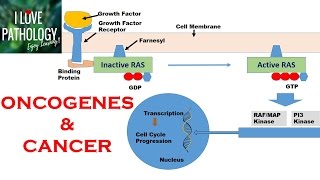
implants that are discontinuous with the primary tumor and located in a remote tissue malignant tumors have the ability to metastasize but benign tumors do not now let's discuss a bit more about cancer all cancers display eight fundamental changes in cell physiology these are called hallmarks of cancer let's discuss about them one by one in detail first one is self-sufficiency in growth signals tumor cells have the capacity to proliferate without external stimuli as a consequence of oncogene activation in our normal cells we have a type of genes called proto-oncogenes the function of these genes is to
produce proteins that regulate normal cell proliferation and differentiation in cancer however proto-oncogenes undergo mutations to produce oncogenes as mutations up regulate the function of oncogenes it is called oncogene activation activated oncogenes produce oncoproteins these oncoproteins have the ability to promote cell growth in the absence of normal growth promoting signals second one is insensitivity to growth inhibitory signals our normal cells also have a type of genes called tumor suppressor genes which encode proteins that regulate cell cycle checkpoints when a dna damaged cell undergoes division these proteins will halt its proliferation and drive the cell to initiate
dna repair if dna repair is not possible they induce cell death by apoptosis in cancer however these tumor suppressor genes are inactivated resulting in uncontrolled proliferation of mutated cells examples for tumor suppressor genes include retinoblastoma gene p53 gene also known as guardian of the genome apc gene wt gene and brca1 and 2 genes i encourage you to find specific types of cancers associated with these genes as an activity third hallmark of cancer is altered cellular metabolism in general glucose uptake of malignant tumor cells is high however they prefer lactic acid fermentation over oxidative physician for
atp generation this is called the warburg effect if glucose has undergone oxidative phosphorylation it produces more energy but all the carbon molecules in glucose are lost as carbon dioxide and even the energy production is less lactic acid fermentation rapidly yields metabolic intermediates that are required for the synthesis of macromolecules essential for the growing tumor this is the main reason that tumor cells prefer lactic acid fermentation as their primary energy production pathway next one is evasion of apoptosis we already know that when a cell with genetic damage tries to undergo cell division it is recognized by
p53 genes and halt its proliferation then the cell is drive towards dna repair and if it is not possible the cell will undergo death by apoptosis this apoptotic process is regulated by another type of genes called apoptosis regulating genes in cancer however these genes are also damaged so there will be accumulation of cells with genetic damage which facilitates development of malignancy fifth hallmark of cancer is their limitless replicative potential also referred to as immortality of cancer cells normal cells in our body can divide only up to 60 to 70 cycles but cancer cells can divide
limitlessly three closely related factors are responsible for the immortality of cancer cells one is evasion of senescence with cellular aging the ability of division in our normal cells declines and the cells become senescent this is thought to be due to the up regulation of p53 gene activity with cellular aging however in cancer p53 gene function is impaired as we discussed earlier so the cells do not reach the state of senescence second mechanism is evasion of mitotic crisis in our chromosomal ends we have specific areas called telomeres these are specific dna sequences that bind with protective
proteins the main function of telomeres is to prevent enzymatic degradation of chromosomal ends and to maintain genomic stability in normal cells telomeres shorten with each division and finally they undergo death due to destruction of dna this is called mitotic crisis however some cells have the ability to re-synthesize telomeres as they have the enzyme telomere race cancer cells also have this ability by doing so they can evade mitotic crisis third mechanism is ability of self-renewal this means that with each division of a stem cell and neoplasia one daughter cell undergoes differentiation and becomes a mature cell
and one daughter cell remains a stem cell so with time there will be more and more stem cells in the neoplasm ensuring that the tumor will not run out of stem cells which are essential to increase the size of the tumor sixth hallmark of cancer is sustained angiogenesis formation of new blood vessels is called angiogenesis like all other cells cancer cells also depend on the body for oxygen and nutrients so they need a good blood supply to maintain their proliferation rate in normal tissues angiogenesis is tightly regulated by a balance between angiogenesis promoting and inhibiting
factors in cancer however this balance is lost maybe due to up regulation of angiogenesis promoting factors or may be due to down regulation of angiogenesis inhibiting factors or may be due to both these factors are secreted by tumor cells macrophages and other stromal cells examples for angiogenesis promoters include vascular endothelial growth factor and basic fibroblast growth factor examples for angiogenesis inhibitors include angiostatin and endostatin seventh hallmark of cancer is ability to invade and metastasize in other words spreading of the tumor to other sites i will discuss about this in more detail towards the end of
this video and the final one is ability to invade host immune response our immune system constantly scan for malignant cells and destroy them this is known as tumor immune surveillance due to mutations malignant tumor cells express abnormal antigens our immune system recognize these as foreign agents so they eliminate these cells by destroying them main cells involved in tumor immune surveillance are t cells macrophages and natural killer cells in immuno-compromised patients tumor surveillance is poor so they are more susceptible to get cancers some tumor cells have the ability to escape the host immune response by several
mechanisms tumors produce large amounts of antigen-negative cells and small amounts of antigenic tumor cells antigen negative cells cannot be recognized by the immune cells so they can easily escape the immune response and some tumor cells secrete cytokines to inhibit immune cells some tumor cells have the ability to mask their antigens for example some cells secrete mucopolysaccharides to cover their antigens and some tumor cells bind to activated platelets in blood and hide their antigens contact dependent killing of immune cells by the tumor cells is another mechanism and some tumors recognize cytokines as growth factors so instead
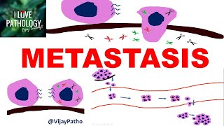
of being killed they undergo proliferation using cytokines now let's discuss about the spread of malignant tumors this topic is extremely important in oncology because invasion of adjacent tissues and metastasis are the major causes of cancer-related morbidity and mortality metastatic cascade is divided into two phases invasion of extracellular matrix and vascular dissemination homing of tumor cells and colonization first let's discuss about invasion of extracellular matrix this is a four-step process but first tumor cells detach from each other due to loosening up of cell to cell interactions then they degrade the extracellular matrix by releasing proteases in
the third step tumor cells attach to novel extracellular matrix components and the final step is migration and invasion of tumor cells in normal tissues cells are tightly bound together by transmembrane glycoproteins belonging to the family of caterins however during invasion of malignant tumors cadherin production is down regulated resulting in detachment of cancer cells from each other as indicated by the yellow circle in this picture in the second step tumor cells secrete proteolytic enzymes to degrade extracellular matrix proteins they also induce surrounding fibroblasts and inflammatory cells to secrete these enzymes main proteolytic enzymes include matrix metalloproteinases
cathepsin d plasminogen activator and collagenases in the third step tumor cells dissociate from the extracellular matrix proteins and bind new extracellular matrix components in normal tissues cells are bound to extracellular matrix proteins like collagen and laminin through integrin receptors this binding helps to keep all the cells in a polarized manner in cancer cells these receptors are down regulated resulting in detachment of tumor cells from extracellular matrix proteins and binding with new extracellular matrix components if normal cells are mobilized from the extracellular matrix like this they undergo death by apoptosis but as tumor cells can escape
apoptosis they survive after detachment from ecm proteins migration and invasion of tumor cells is a multi-step process involving many types of receptors and signaling molecules cleavage products of matrix proteins and growth factors such as insulin like growth factor act as chemotactic agents and facilitate the amyloid movement of cancer cells finally they make their way into the vascular system in order to metastasize this is called intravasation next phase of metastatic cascade is vascular dissemination homing of tumor cells and colonization metastasis is the process of producing tumor deposits at a site distant to the initial mass and
these secondary deposits are discontinuous with the primary tumor metastasis occurs via three pathways seeding of body cavities and surfaces lymphatic spread and hematogenous spread seeding of body cavities and surfaces occurs when the tumor directly penetrates into a natural open field these areas lack physical barriers one common example is peritoneal cavity lymphatic spread is the most common way for the spread of carcinomas but some sarcomas may also disseminate via lymphatics hematogenous spread is the most common way for the spread of sarcomas arteries are usually resistant to invasion by tumors because they have a thick wall and
higher pressure inside so most tumors prefer veins over arteries once in the circulation after intravasation tumor cells are vulnerable to destruction by several mechanisms including mechanical shear stress innate and adaptive immune defenses and apoptosis induced by mobilization from the extracellular matrix so tumor cells tend to clump together in the circulation as an adaptive response to escape these mechanisms they also bind activated platelets and coagulation factors this whole structure is called a tumor and bolus formation of tumor and bully helps tumor cells to withstand shear stresses and escape the immune response in the circulation once a
favorable soil is reached tumor cells then extravasate from the circulation in order to produce metastatic deposits the site of metastasis is determined by natural pathway of venous and lymphatic drainage and organ trophism of tumor cells finally let's discuss about some common clinical consequences due to the spread of malignant tumors first let's take local effects these effects can be categorized into three groups effects due to the tumor mass effects due to local regional spread and local hormonal effects these effects of local invasion are dependent upon the site of the tumor compression on the normal tissue and
adjacent structures by the tumor may cause loss of function of the affected area for example tumors in brain tissue may cause certain neurological defects and herniation of some part of the brain as tumors have a high proliferation rate some cells may not receive adequate oxygen to maintain their functions so they may undergo necrosis this will result in ulceration of the affected area for example in colon cancer mucosa of the colon frequently get ulcerated due to necrosis some tumors act as a nitis for infection for example some people develop bacterial pneumonia and lung cancer when the
tumor involves a lumen of an organ it may cause luminal obstruction for example in colon cancer lumen of the colon gets obstructed due to the tumor mass another manifestation of malignant tumors is perforation of the organs for example malignant perforation of gastric ulcers and this may result in peritonitis ultimately local hormonal effects occur when the tumor is a hormone secreting one this may result in excess hormone levels in blood effects due to local regional spread may occur when the tumor erodes into surrounding structures like veins nerves and body cavities for example invasion of veins may
cause varicocele in patients with renal cancer invasion of nerves may cause nerve palsies and invasion of adjacent body cavities may cause effusions most of the time cancers cause systemic manifestations as well cancer cachexia is one of the most common manifestations in cancer that occurs in about 50 patients with cancer it is characterized by the equal loss of both fat and lean muscle elevated basal metabolic rate and evidence of systemic infection tumor necrosis factor is the main mediator responsible for cancer cachexia inhibitory effect of tumor necrosis factor on the hypothalamus causes anorexia and many patients will
have increased levels of acute phase proteins paraneoplastic syndromes are a group of disorders that are triggered by an abnormal immune response against malignant tumors occur in about 10 to 15 percent of patients with cancer patients with paraneoplastic syndromes will have a wide variety of symptoms and diseases including hypercalcemia hypoglycemia polycythemia disseminated intravascular coagulation finger clubbing cushing syndrome acanthosis negligence and nephritic syndrome okay that's all i wish to discuss in this video i have covered most of the learning material required for general pathology on neoplasia but do not forget to read your textbooks there is another
topic in neoplasia the molecular basis of cancer i will discuss about it in another video
Related Videos

12:36
What is Cancer ♋️ ? What is Tumor (Neoplas...
Medicosis Perfectionalis
93,515 views

54:56
Neoplasia (Part 1) : Definition, Nomenclat...
Rabiul Haque
375,318 views

14:43
NEOPLASIA-1: BASICS: Nomenclature: Benign,...
ilovepathology
275,192 views

16:06
Why do people get cancer, how it spreads, ...
TEDx Talks
1,422,059 views

10:44
Chronic Inflammation : Causes, Morphologic...
Med Today
129,783 views
48:33
Introduction to Cancer
Aaron Mullally
254,151 views

8:02
1. Neoplasia part 1: definition, how it re...
Oncology for Medical Students
169,988 views
22:13
Cell Death : Necrosis & Apoptosis - Types,...
Med Today
96,191 views

1:35:03
Lung Cancer
Ninja Nerd
97,464 views

9:19
NEOPLASIA Part 11: GRADING and STAGING of ...
ilovepathology
44,721 views

25:32
Benign Prostatic Hyperplasia vs Prostate C...
Dr Matt & Dr Mike
137,890 views

36:00
Hematology | Types of Anemias
Ninja Nerd
2,793,518 views

10:10
Hallmarks of Cancer | Pathophysiology
Dr Matt & Dr Mike
50,881 views

12:50
Cell Adaptations : Pathology - Hypertrop...
Med Today
180,652 views
17:55
NEOPLASIA 9: TUMOR IMMUNITY: tumor antigen...
ilovepathology
63,095 views
9:18
Wound Healing - Stages of healing and path...
Armando Hasudungan
149,424 views

8:44
2. Neoplasia part 2: Differences between b...
Oncology for Medical Students
150,267 views
14:25
NEOPLASIA 8: INVASION AND METASTASIS; Mech...
ilovepathology
139,341 views
10:37
Understanding Pancreatic Cancer
Zero To Finals
254,556 views
10:39
NEOPLASIA 2: HALLMARKS OF CANCER : Protoon...
ilovepathology
189,981 views