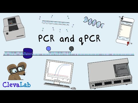
What is PCR and qPCR? | PCR Animation
200.92k views1048 WordsCopy TextShare
ClevaLab
Discover molecular biology and the Polymerase Chain Reaction (PCR). What is PCR and qPCR? PCR takes ...
Video Transcript:
ClevaLab, Basic Principles of Human Biology Explained. An American scientist, Kary Mullis, invented PCR in 1983. His idea was to use two primers, nucleotides and DNA polymerase, to make unlimited copies of DNA.
A primer is a 20 base pair fragment of DNA that pairs the target DNA. The DNA sequence of the primers is so specific that from a background of 22,000 genes they can amplify one gene. DNA exists as two strands bound together.
So for a primer to access the DNA, the strands first need to be separated by heating at 95 degrees Celsius. As the mixture cools to 60 degrees, the primers bind to opposite strands of the DNA. Binding so they flank the target region.
DNA polymerase can only copy DNA in one direction. So the strands get made in opposite directions resulting in overlapping copies. This copying of the DNA by DNA polymerase will double the amount of DNA.
The reaction is again heated to 95 degrees to separate the DNA. When cooled, the primers also bind to the new DNA, and the DNA polymerase makes another copy. For each PCR cycle, the amount of DNA doubles.
So that two copies of DNA can become two million copies in just 20 PCR cycles. The flanking placement of the primers limits the length of the amplified DNA. This DNA can be visualised on a gel at the end of the amplification and will be one discrete band.
Heating the mixture to 95 degrees destroys DNA polymerase. So fresh DNA polymerase gets added after each heating cycle. This manual addition was very time consuming and error-prone.
But in 1988, Randall Saiki, from Cetus Corporation, used a different DNA polymerase for PCR. One from Thermus aquaticus or Taq. Thermus aquaticus is a bacteria that lives in hot springs.
So it isn't destroyed during the 95-degree heating step. The use of taq polymerase means that PCR can continue to the end without having to add more polymerase. Cetus also created an instrument to automate PCR.
The first thermal cycler, the TC1, automated heating and cooling using a metal heat block and an inbuilt computer. This automation made the process of PCR far easier. The first commercial real-time PCR instrument was made in 1996 by Applied Biosystems.
This instrument monitors fluorescence in the PCR tube as it is happening. DNA production is measured using two primers and a probe. A fluorescent dye is attached to one end of the probe, and a quenching dye is on the other end.
When the probe is intact, the quencher stops the fluorescence. The probe sits close to one of the primers. As Taq polymerase copies the DNA, it cuts up the probe and releases the dye and quencher.
The quencher is no longer near enough to stop the fluorescence, so light gets emitted. The camera then records the fluorescence. The increase in DNA during PCR can be seen on the computer screen in real-time as the PCR is cycling.
The power of real-time PCR, also known as quantitative PCR or qPCR, is to see when the PCR is doubling each cycle without stopping the reaction. In this doubling phase, the quantification of DNA is possible. In the first few cycles of the qPCR, the amount of fluorescence is below the camera's detection limit.
Then, as DNA accumulates, the fluorescence in the tube becomes detectable over the background. At this point, the fluorescence doubles each cycle. This doubling is the exponential growth phase.
As millions of copies of DNA accumulate, the reaction slows down to a linear rate and then reaches a plateau. The higher the starting amount of DNA, the earlier the PCR will appear above the background. The amount of DNA in different samples is measured using a threshold line.
The point that the fluorescence passes this threshold is the cycle threshold or Ct of the PCR. The threshold is set above the background and during the exponential phase of the PCR. The Ct value is a measure of how much DNA is in the sample.
But, the Ct value can't tell you the exact amount of DNA in the tube. It can only tell you a relative amount. For example, a sample with a Ct of 10 will have two times more DNA than a sample with a Ct of 11.
That means that a difference of two cycles is four times more DNA, and 20 cycles is a million times more DNA! So, if you want to know the exact copies of DNA in a tube, you need to compare it to a known amount called a standard. Standards covering the full range of qPCR cycles can make a standard curve.
Then when you know the Ct of a sample, you can read the DNA copies from the standard curve. It's also possible to amplify more than one DNA target in the same tube. Each target uses a probe with a distinct coloured dye.
Each primer and probe set can target different locations in the DNA or even slight changes in the sequence. This change can be as small as one nucleotide in the DNA sequence. PCR and real-time PCR have many uses.
For example, in diagnosing disease and in medical research. Top of mind is the detection of viruses and bacteria from respiratory infections. Most of us have had a swab or given a saliva sample for one of these tests.
From this sample, PCR is used to identify which viruses or bacteria are present. Different coloured probes in one PCR reaction allow for the detection of many targets in one PCR. For example, it's common for one test to detect influenza A, influenza B and SARS-CoV-2.
These are yes/no tests, meaning they report if a virus or bacteria is detected or not. But for COVID-19, the Ct value of the real-time PCR is also helpful for contact tracing. A low Ct means that the person has a lot of virus present and is more likely to be infectious.
Tests over time can tell if the person is early or late in their infection and when they were infectious. The discovery of PCR was a fantastic insight. PCR enables Disease Diagnosis and a greater understanding of biology.
Subscribe to ClevaLab to learn more about human biology.
Related Videos

8:03
What are Autoimmune Diseases and How Do Th...
ClevaLab
236,669 views

12:36
What is Quantitative PCR?
Osmosis from Elsevier
13,716 views
14:12
qPCR details | quantitative real time PCR ...
Animated biology With arpan
84,624 views

14:37
Sanger DNA Sequencing, From Then to Now.
ClevaLab
77,607 views

10:49
PCR (Polymerase Chain Reaction) Explained
BOGObiology
104,749 views

12:40
Western Blot Protocol
SENS Research Foundation
61,740 views

3:49
SYBR Green vs TaqMan – How qPCR works
INTEGRA Biosciences
42,765 views

7:31
What is Gel Electrophoresis? | miniPCR bio™
miniPCR bio
413,353 views

8:20
Coronavirus Test: Real time RT-PCR - Anima...
Biology with Animations
5,543,154 views

21:25
PCR Basics
Joseph Ross
144,627 views
4:44
Next Generation Sequencing (Illumina) - An...
Henrik's Lab
640,996 views

58:07
Real-Time PCR in Action
USDA Animal and Plant Health Inspection Service
456,611 views

lofi hip hop radio 📚 beats to relax/study to
Lofi Girl

6:41
Founding Fathers Cold Open - SNL
Saturday Night Live
3,731,947 views

53:33
Your Brain: Who's in Control? | Full Docum...
NOVA PBS Official
4,973,760 views

36:12
Understanding PCR
Mark Temple
82,537 views

6:10
What is RT-PCR? (Real-Time PCR & Reverse T...
YouTooBio
82,691 views

37:26
Molecular animation – Tech Talk by Drew Be...
WEHImovies
83,330 views

7:55
Gel Electrophoresis
Amoeba Sisters
2,404,149 views
31:26
Next Generation Sequencing 1: Overview - E...
iBiology Techniques
480,537 views