EASY TO UNDERSTAND | Introduction to Nervous System
239.54k views3336 WordsCopy TextShare
Miss Angler
Join this channel to get access to perks:
https://www.youtube.com/channel/UCjA2nEpHzkvVjROX-rqzdzg/j...
Video Transcript:
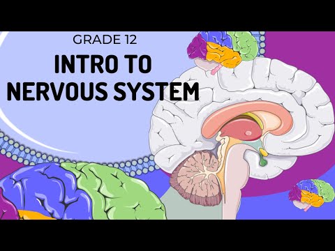
hi everybody and welcome back to miss angler's biology class i am miss angler and in today's video we are going to look at the nervous system this is an introductory video so i'm going to run you through the basics the functions how to identify the different structures and most importantly i'm going to run through the things you need to know for your exam now if you are new here don't forget to give this a video a thumbs up and subscribe and make sure your notifications are turned on because i post new videos every tuesday and
thursday now starting off with the basics you are going to need to know what exactly is the function of the nervous system and the function of the nervous system can be summarized into a very simple flow diagram and i'd like to also point out that you can use this flow diagram to explain any response that happens in the nervous system anything about to draw for you is you can use this also to explain your endocrine system as well so in any kind of system where there is a response to some kind of external environment we
always start our story with some kind of stimulus now a stimulus in the nervous system could be something like light it could be pain it could be sound and whatever the stimulus is it needs to be received by someone so that incoming stimulus is going to be sensed to some kind of sensory receptor and that is generally a century set of cells maybe in the eye in the ear in the skin they receive that information now after that that information needs to be sent to someone and in this diagram we are only going to focus
on the central nervous system and the central nervous system is responsible for taking in this information and it needs to make a decision and so when we speak about these decisions from the central nervous system we call it its integrative function in other words your central nervous system decides what's going to happen next so your brain and your spinal cord make a decision are we going to blink are we going to run away what are we going to do now once we've made that decision we need to send that decision to somebody who is going
to carry out the response and so we call these structures effectors they are generally muscles or glands and they respond to this instruction and they follow out and they create some kind of response which is our final portion of our flow diagram here responses can be things like increasing the rate of digestion or maybe decreasing it maybe it increases your heart rate and so effectively in that example the effector would be the heart and increasing the heart rate would be the response and so this is how you would explain how the nervous system functions in
order to bring about a change within the body due to some kind of external stimulus now let's get into the structures that make up the central nervous system and when i speak about the central nervous system i'm speaking about the brain and the spinal cord now sitting around the brain and spinal cord are structures called meninges which are protective layers that sit around the brain now this is a little cross section through a piece of brain as well as some protective layers so this gray area over here represents the brain on either side and i'm
going to start off with the outside layers first so this outer layer that we see over here is actually your skin then sitting just below that is the bone of the skull and then there are three protective layers that sit underneath that in order to protect your brain as well as your spinal cord we start with the most outer layer which is this thick tough blue layer it is called the dura mater it's very fibrous it's almost like a sac that your brain sits in then sitting just below that is this area over here which
is in the sort of paler blue in the middle that is the arachnoid layer it is where all your blood vessels are found and then the most inner meninge which sits right up against the actual brain itself it's a very delicate layer is called the pia mata now the pia mata is a very delicate layer and all together the meninges are protective layers around both the spinal cord as well as the brain and so when we talk about the function of the meninges we say they are there for protection and they also allow for cerebral
spinal fluid to be secreted and cerebrospinal fluid assists with lubrication but also with protection making sure that your brain is well secured we're starting off with the main component of your central nervous system which is the brain and the brain has regions to it and first of all we're going to start off with the largest part of your brain which is this folded area on the outside of your which is called the cerebrum and the cerebrum is essentially where you live in the sense that it's your personality it's your memories and all of those folds
and grooves are called silky and diary and essentially that is where all of your sight comes from your hearing your memories everything is housed in this part of your brain and so when we list the functions of the cerebrum we say it's where voluntary movement comes from so like walking and exercising and moving around it's also where your senses are as i mentioned and most importantly your cognitive function it's where your intelligence is then sitting at the bottom or the lower underside of your brain is this a gray area at the back here this is
called the cerebellum now the cerebellum is quite an old region of your brain and in different animals of different sizes um and that is something to do with the complexity of the animal our cerebellum is a little bit smaller because we have such a large cerebrum but the function of the cerebellum is it coordinates your voluntary movement in other words if you want to walk nice and smoothly um the cerebellum is responsible for that and make sure that you can walk in a nice straight line and that it's a smooth movement that's not jerky it'll
maintain general tone now the stance doesn't mean your muscles look good or toned what it means is that your muscles have some firmness to it when you're awake so that your muscles can keep you upright and stable and that links into the last function which is posture and balance your cerebellum plays a really important role for understanding where is your head in relation to your body when you're moving but also when you are moving and you're walking to keep you in a straight line the cerebellum make sure that you continue to do that now in
order to see the rest of the components of the brain we're going to have to cut it open so we can see on the inside and here is a lovely cross section through the cerebellum at the bottom here as well as seeing a cross section through the cerebrum on the top but the next structure i want to bring attention to is this pink structure over here this is called the corpus callosum now the brain is actually divided into two hemispheres or two halves a left and a right hemisphere each hemisphere controls the opposite side of
your body so the left controls your right and your right controls your left and the function of the corpus callosum is to make sure that there is good communication between these two hemispheres it ensures that the left hand side of your body knows what the right hand side of your body is doing then we're going to move in to this lower region of your brain reaching down in to what we call the medulla now the medulla is this swollen region at the bottom it's the medulla oblongata and it has a really important function because this
is where life actually is sustained in your brain and you'll see now as we look through the functions the functions include things like the pathway for impulses from your whole body your reflex center for breathing and heart rate and so many more things as well as less important reflexes like sneezing and coughing if your modula is damaged in any way it can often lead to your death you don't breathe you can't you know your heart doesn't beat and so it's a really important part of the brain often referred to as part of the brain stem
and we can call it a stem because if you look at it this region from about here to here does look like a stem of a flower now some people in some textbooks are not so sure about when you call it the medulla oblongata and what do you call it the spinal cord well you call it the medulla as long as you are still inside of the cranium in other words you haven't left the skull bone but the moment you leave the skull bone and let's say that's over here that was the exit out of
the skull everything beyond that and below that is called the spinal cord and a nice way to also figure that out is there is a large hole in the bottom of the cranium called the foramen magnum it is where the spinal cord leaves the brain and so that is the beginning of the spinal cord if you were going backwards and upwards it would then go from the spinal cord and then after the foramen it would become the medulla oblongata let's move into the spinal cord which is the second component of your central nervous system and
it's technically like the tail of the brain it's completely attached to the brain it is it is a part of the brain and it extends all the way down your vertebral column and we've cut the spinal cord in half so you can see on the inside here and i'm going to start with the differences in color that you'll notice you'll see that on the inner region of your spinal cord the matter is actually darker so we call it gray matter versus the outer region of your spinal cord is white matter and the difference in color
is linked to something called myelin and i will get back to that at a later video it is a fatty substance that sits around our nervous tissue and so the more you have of it um the change in color will occur now i'd like you to also know that when you're looking at a spinal cord like you are here on the right hand side so on this side over here we have some spinal nerves and these are nerves that are going to leave the spine and go to the rest of the body i'd like you
to know that there is also going to be one on this side but for the simplicity of this video i have just left it off so that it doesn't influence how we label the diagram but it's technically on both sides and so you're going to need to be able to label this particular diagram and i'm going to start off with what these structures are over here now this upper region over here where there's a bit of a bulge is called the dorsal root dorsal meaning back in other words that is the back of your spinal
cord it is the part that is closest to your skin or where your vertebral column is and then the other or opposite side to that if that is the dorsal side is what we call the ventral root i'm also going to tell you why you need to know these slightly later in the video why it's important as well for later videos the hole in the center over here is called the central canal and there is some cerebrospinal fluid in there as well and then the final thing is this structure coming out of our spinal cord
which is the spinal nerve now when we speak about the spinal cord's functions we know that it is a pathway of imp for impulses it is where primitive reflexes exist and when we talk about a primitive reflex we're talking like the knee-jerk reaction or the pain response when you pull away from something that is painful now the reason why we need to know the structure of the spinal cord is because in a later video i'm going to explain a reflex arc but just the basics for now so you can place it when a pathway of
impulse moves from your spinal nerve into your spinal cord it is going to travel along the spinal nerve it is going to go into the dorsal root and down into the spinal cord itself a decision is then made in the spinal cord and that's why it's called a reflex so it actually completely just jumps the brain and then the decision that is made leaves out the ventral root and then back down the spinal nerve once more and this is what we call a reflex arc i'm going to do that in a lot more detail in
a later video now that we've covered the central nervous system which as you can see in this picture over here is the brain and the spinal cord we now need to move into the peripheral nervous system peripheral comes from the word like on the periphery like on the edges and the peripheral nervous system is all the other nerves of the body it is everything that runs down into your arms and your legs and to all the organs of the body and that is divided into two types of systems again we have the autonomic nervous system
and we have the somatic nervous system now we'll start with the somatic because it's the easier one and it doesn't need a lot of explanation somatic means body and it refers to the nervous system that is communicating with your sense organs and your voluntary muscles and so the somatic nervous system would be the one where you would be like moving your eyes to look somewhere you would also be walking and so this touches on the sensory part of it as well as the motor which is the movement part what we're going to focus in more
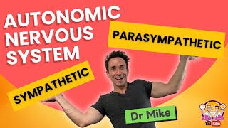
detail is the autonomic nervous system and this communicates with all the systems that are automatic in other words you don't need to think about it they're involuntary they just happen and you'll see now why it's important for them to just happen without you thinking about it and the autonomic nervous system divides again into two and it's called the sympathetic division or the parasympathetic division now looking at this in the diagram we're going to be able to apply what it actually means when we talk about the autonomic nervous system and it's two divisions and i'm going
to start on the right hand side with the sympathetic nervous system all of the organs of the body as long as as well as some of the glands are double innervated it means they've got two nerves going into them and essentially the reason why they have that is you need to be able to control how they are reacting and so on the sympathetic side of this system this is where we have the flight or fight response and so if you read some of the descriptors that go down the sympathetic nervous system if you are running
away or scared of something your pupils dilate your heartbeat increases um you have an increased rate of glycogen to glucose because you need to access sugar because you need to run away um and there's a decreased activity in your digestive system you don't want to waste any energy you'll also notice that if you look at the picture there are direct links coming out of the spinal cord to each of these organs and that's because the sympathetic nervous system needs to be automatic quick fast get a response on the other side however the parasympathetic nervous system
and the word power should give it away it's more relaxing and this side we say it is a rest and digest system and it allows your body to do just that and you will notice that it has all the opposite functions that we saw in the sympathetic it causes your pupils to constrict and let less light in um it slows your heart rate um and essentially the parasympathetic side is when your body is not under any stress and it's just relaxing and these two systems are completely automatic they are run by your brain you don't
have to think about them they are involuntary and most importantly you also notice that there is a single nerve that is running out to most of these and that is because when you are resting and digesting you don't need a lot of electrical impulses to go to these structures once you tell them what to do they keep doing it and they'll do it very slowly over time whereas the sympathetic needs a specific nerve going to each of these regions because we want a fast react reaction now as always i like to finish off my lessons
with a terminology recap now there was a lot of terms in this section and there's going to be even more as we go through the eye and the ear as well so i'm going to be very brief but you can use these words to create flash cards it's a great way to study for biology so we spoke about stimulus which is an incoming message from the external world like sound or pain or heat there's a receptor organ of some kind receiving that information the integrative function is your brain making a decision that decision is set
to a effect or organ like a muscle gland and then we went into the actual central nervous system which is made out of the brain the brain is covered in meninges and they are the pia mata the arachnoid and the dura mater we then looked at the regions of the brain which is the cerebrum the corpus callosum cerebellum medulla oblongata and then the final component of the central nervous system was the spinal cord we then looked at how besides the central nervous system the other divisions of the nervous system which is the peripheral nervous system
that peripheral nervous system is all the other nerves of the body it then divided into the somatic and then the autonomic nervous system the autonomic nervous system divided again to become the sympathetic or the parasympathetic nervous system it's quite a lot to wrap your head around so i suggest drawing some kind of almost diagram of a family tree or sort of a flow diagram to see how they split from one another a very similar one that we saw in two pictures back where it had organized it very well in that flow diagram now as always
if you've liked this video don't forget to give it a thumbs up and subscribe and turn your notifications on because i post every tuesday and thursday and i will see you all again soon bye
Related Videos

17:54
Nervous system Introduction
Miss Angler
81,575 views

7:43
Easy to understand | REFLEX ARC
Miss Angler
101,962 views

12:20
INTRO to the Structure of the EAR (UPDATED...
Miss Angler
8,937 views

23:04
How Ultra-Processed Bread Took Over Americ...
Business Insider
212,822 views

10:47
Human Nervous System (Part 2) - How the Br...
Thomas Schwenke
760,191 views

19:05
Nervous system anatomy introduction
Sam Webster
94,026 views
39:23
Autonomic Nervous System (Sympathetic & Pa...
Dr Matt & Dr Mike
230,517 views

23:34
Why Democracy Is Mathematically Impossible
Veritasium
3,616,248 views
10:36
The Nervous System, Part 1: Crash Course A...
CrashCourse
9,340,994 views

12:28
INTRO to Accommodation of the EYE (UPDATED...
Miss Angler
7,597 views

5:09
3 tips on how to study effectively
TED-Ed
4,125,374 views

11:53
Biggest Breakthroughs in Biology and Neuro...
Quanta Magazine
852,568 views
29:43
The Surgery That Proved There Is No Free Will
Joe Scott
1,603,233 views
14:47
Neuroanatomy S1 E1: Intro to the Central N...
UBC Medicine - Educational Media
2,098,316 views
21:10
How to Slow Aging (and even reverse it)
Veritasium
7,259,720 views

13:56
The Brain
Bozeman Science
5,801,397 views
10:15
Nervous system (central & peripheral) | Co...
Khan Academy India - English
331,217 views
9:22
The Nervous System In 9 Minutes
CTE Skills.com
7,607,689 views

26:54
How Your Brain Organizes Information
Artem Kirsanov
541,429 views

17:02
Somatic vs Autonomic Nervous System | Phys...
Medicosis Perfectionalis
83,432 views