🥇 CAVIDAD BUCAL, LABIOS y MEJILLAS. -Anatomía- ¡Explicación Sencilla!
187.13k views2431 WordsCopy TextShare
Anatomía Fácil por Juan José Sánchez
Descarga esta y cualquier diapositiva de mi canal, uniéndote a PATREON!
También puedes unirte a los ...
Video Transcript:
hi how are things? How are you doing? Welcome to a new easy anatomy video by Juan José Sánchez.
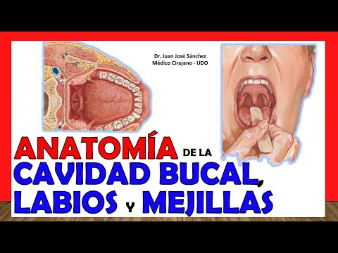
Today I bring you a video about the anatomy of the oral cavity or oral cavity as you want to call it and we will take advantage of this topic since it is relatively short, well we will include the anatomy of the lips and cheeks, which is known as the of the cheek, here we are going to talk about the generalities of the mouth, there is a separate video in which I will explain the palate, the anatomy of the palate can be found on my channel that I will publish in parallel with this one on the oral cavity and they will also find a video about the tongue, which is a really long organ, a visor that is long to explain and that is why I dedicated only one video to explaining it. So this oral cavity is also called the oral cavity, you should not confuse the con vowel with a small v because that refers to the vocal cords of the voice, something that is in the larynx, here it is buccal with a high b that refers to the cavity from the mouth; So this oral cavity will be comprised of two parts: a more external part, which is actually smaller, is called the vestibule, which is what continues after the lips, when you close your mouth. He closes his teeth, glues the upper ones to the lower ones, everything that is left outside the teeth between the teeth and on the lips, that is the vestibule, it has to be a small cavity, some books say that it is a virtual cavity, Now everything that is left inside me is enclosed by my teeth where the tongue is, where the palate is, that is the second part of the oral cavity.
Look at this image. What you see in green is everything that is, let's say between the teeth, when I close my mouth is what is left inside my teeth, what I cannot see when I close my mouth, when I close my teeth better said that is the famous oral cavity itself; Now everything that is left on the outside, the basis that we should study this is with a closed mouth but well I did not find an image that I am going to explain but more or less you understand that everything that is outside the teeth is the vestibule or entrance and everything that remains inside my mouth when I close my mouth that is the oral cavity itself, we are going to see other images to see if they understand it a little better and we are going to explain in more detail the anatomy of each of them. these portions of the oral cavity, but first do not go away from the video, it is important that you subscribe to the channel, I remind you below where it says subscribe, you click on it and you are automatically subscribed to the channel and you can access the more than 200 anatomical videos that are there, if there is any video that you do not see within the content of the channel, you can write it to me in the comments.
Good thing, I take it into account and I always watch them when making the following videos. So we start first talking about the vestibule, notice that the external limit of the vestibule is going to be the lips and the cheeks are going to be what we know colloquially as the cheek, we are going to see it better that would be the external limit of the vestibule, while that the internal limit is going to be the teeth and the gums are going to be, that is what is going to internally separate that vestibule from the oral cavity itself, also notice that this is a previous view with the palette we open a little plus the mouth opening to be able to see what the lateral wall of that vestibule is like and we see that we precisely find the area of the cheek, there is a very important structure that we find in relation to that cheek, it is this hole called the parotid papilla, they also call it caruncula . parotid duct, that orifice is the place where the parotid duct or also called stenon duct empties and following this parotid duct then carries the salivary secretion from the parotid gland to, let's say, inside the oral cavity, but you already know that it specifically empties into the vestibular portion .
How do we locate this parotid papilla? We are located on the upper second molar and there we are going to find that hole, all people have it, look in the mirror so you can see that the parotid papilla is there, we are also going to see that it exists, so you close your teeth, make an occlusion, there is a communication between the vestibule, which is what you can see here perfectly, and the oral cavity itself, and that communication is behind the last molar, If the person already had a good third molar, it will be behind the third molar or, if not, the second molar will be the most posterior one, and that will be between that molar and behind the ascending branch of the jaw, which is this little bone that It is here, the ascending branch of the lower jaw; then that space communicates the oral cavity itself and the vestibule region as such, here you can see the separation between both portions of the oral cavity very perfectly, look here too, here you can see the vestibule area, well, it is half achieved see here and inside the entire famous oral cavity speaking then of the parotid duct and it was pointed out as the parotid papilla but what I want to tell you is that we see the parotid duct as it curves in front of the masseter, it pierces the buccinator muscle that we are going to see right now and it ends right next to the upper second molar, notice, then speaking of the oral cavity itself we are going to see that its anterolateral limit is going to be the same internal limit of the vestibule, practically alveolars that are structures of the lower jaw and the upper jaw, we see both the lower and upper teeth and the gums, it will then be the anterolateral limit of the oral cavity itself, which in fact is incidentally what separates me from that oral cavity itself of the vestibular area, as a posterior limit we are going to find a structure called the oropharyngeal isthmus. Here it is important that you more or less know the anatomy of the pharynx because in that video, by the way, I explained what the oropharyngeal isthmus is like, which is made up of both pillars .
anteriors of the palate also called palatoglossal arches downwards by the root of the tongue and upwards by the soft palate which right now we are going to see a little better, then that circular space called oropharyngeal isthmus is the posterior limit of the oral cavity because behind of that oropharyngeal isthmus is the pharynx, specifically what we call oropharynx or oropharynx but see that it is a truly virtual space and in fact see that part of my tongue remains inside the oral cavity itself, but part of the tongue is part really about the oropharynx, as I repeat, I explain this oropharyngeal isthmus much better in the pharynx video. Notice, there I am pointing out the oropharyngeal isthmus. It is that space.
Here we see the anterior pillars, which are the palatoglossal arches, or if we see above the soft palate, including the uvula as I finish delimiting that same buccopharyngeal below, you already know what is the dorsal part of the tongue part of the dorsal part of the tongue, excuse the cacophony, the roof for its part of the oral cavity itself the palate As I tell you in a separate video, I explain the anatomy of that palate because it is a very extensive anatomy to include in this oral cavity video. We are going to see that the floor is also going to be the tongue and I also explained to them that there was a video about the tongue, a single explanation. We are going to see then that under the tongue, we continue with that floor of the oral cavity, we find what we call the floor of the tongue.
the mouth, which is made up mainly of the famous oral diaphragm, which is the mylohyoid muscles, as I am pointing out there, we are going to see that when we raise the tongue, that lower surface of the tongue shows certain anatomical structures, we are going to see first in the midline a structure that joins it to the floor of the mouth, I am talking about the tongue, which would be the frenulum of the tongue. On each side of the frenulum of the tongue, we are going to see some ducts, the mouth, that is, one on each side, which are the caruncles. sublingual, also called sublingual papillae, which is where the sublingual duct, which is the Wharton duct, opens, ok?
Wharton's duct, also well, these papillae are not really the sublingual one, this one that I am pointing out to you, this is the submandibular papilla, that is where the Wharton's duct empties, I correct, well you already know that we are from the family, that sublingual here is not really sub maxillary because there the maxillary gland empties through the Wharton duct also called submandibular duct, it is one on each side of the frenulum of the tongue, let's see if we Going to the sides we see some elevations that are called the sublingual folds because it is the part of the mucosa that is above the sublingual gland. Likewise, we will find at that level many small ducts that are the ducts or the places where they empty. the sublingual ducts, then you know that these are the sublingual ducts but this is the submandibular papilla, here there was actually a transcription error between sublingual and submandibular, those structures are found within the oral cavity itself.
Let's now move on to the anatomy of the lips, remember that we actually had two lips, we talk about an upper lip and a lower lip. They are structures quite filled with connective tissue but they hide inside them the muscles or the orbicularis oris muscle. You see here that I am pointing out this muscle was very well explained in my video on muscles of facial expression, very well let's see then how I tell you that we have lips, an upper lip, an upper lip and a lower lip, both Lips join in a region in a corner called the labial commissure, the labial commissure we are also going to see a structure that we are going to find in the anterior part of the skin of the upper lip, which is a hollow, a depression that we have there, that depression is called the filter or the philtrum so it is a structure related to these lips, we are also going to see that both the upper lip and the lower lip have a fold that joins it to the corresponding gum.
We call those folds the lip frenulum since The frenum of the upper lip and this is the frenulum of the lower lip are structures that are found inside the vestibule, by the way so as not to confuse, notice something here also that I did not tell you, that the entire oral cavity is really a mucosa. which covers then. See how the mucosa that lines the inner side of the lip continues, as we say, it folds over the gum and into the same mucosa and finishes waterproofing that cavity, that vestibule specifically, we are going to see that the cheeks, for their part, are going to be made up mainly of the buccinator muscle, which is this muscle that you see there, remember that I told you that the buccinator muscle was perforated by the parotid duct so that saliva could reach the parotid gland and into that vestibule, also on those cheeks we found a group of glands that are minor salivary glands, if a set of glands that also produce saliva and we can find many ducts but they are really almost microscopic, imperceptible along the entire mucosa of that cheek, we are going to see that also in relation to this buccinator and in relation to this cheek we find a buccal fat ball that is the famous bichat fat ball, the one that women remove when they have a bichectomy to look more silhouetted at the level of the face, ok?
We are going to see that the union between the upper lip and the cheek region is given by a groove that we see externally, it is a little more marked in some people than others, that groove there from the lateral edges of the nose to the edges lateral corners of the lips, then since it is joining the lip with the nose, it is called the famous nasolabial fold, so hated from an aesthetic point of view by some women, we also see that at the level of those cheeks and that these parts that we see here are going to be find here in the depths the buccinator muscle and that buccinator muscle ends in this raphe called the mandibular pterygo raphe or maxillary pterygo that is separating it from the muscle that is behind it in the superior constrictor muscle of the pharynx, it is said that it ends there from the deep point of view that part of the buccal vestibule and was indicated by the mandibular pterygo raphe.
Related Videos

27:32
🥇 ANATOMY OF THE TONGUE. Easy and Simple ...
Anatomía Fácil por Juan José Sánchez
201,184 views
![La CAVIDAD BUCAL - Sus partes y características [El procesamiento inicial del alimento]](https://img.youtube.com/vi/J2iAJeATb6Y/mqdefault.jpg)
16:36
La CAVIDAD BUCAL - Sus partes y caracterís...
Nutrimente
9,509 views

24:50
🥇 ANATOMÍA DEL PALADAR. -Músculos, Inerva...
Anatomía Fácil por Juan José Sánchez
93,602 views

23:37
Esto le pasa a tu cuerpo al perder las mue...
Dr. Federico Baena Q
1,394,504 views

32:42
Anatomía dental - clase magistral virtual
Prótesis Dentales Angie Lindo Costa Rica
65,067 views

21:51
🥇 NASAL CAVITY 1. (1/2). Easy and Simple ...
Anatomía Fácil por Juan José Sánchez
181,952 views

16:49
🥇HOW MUCH DO YOU KNOW ABOUT THE BONE SYST...
Anatomía Fácil por Juan José Sánchez
511,288 views

18:30
Anatomía de Cabeza y Cuello - Boca y lengua
Medizi
13,446 views

24:30
🥇 ANATOMY OF THE NOSE. Easy and Simple Ex...
Anatomía Fácil por Juan José Sánchez
153,601 views

14:33
FISIOLOGIA de la CAVIDAD BUCAL, MASTICACIO...
david vargas
83,951 views

20:16
🥇 ANATOMY OF THE PHARYNX 1/2, (Muscles, I...
Anatomía Fácil por Juan José Sánchez
229,716 views

14:55
Anatomía de la cavidad oral
El bolígrafo de Juan y el lápiz de Fernando
35,715 views

19:30
🥇 KIDNEY Anatomy (1/2). Easy Explanation!
Anatomía Fácil por Juan José Sánchez
461,327 views

18:33
E1.1 | Boca y faringe | Anatomía 2 | FMed UBA
Anato2ok FMed
27,312 views

24:57
GIRL BULLIED At School For Her MUSCLES | D...
Dhar Mann Studios
1,982,923 views

26:02
Aparato digestivo. Cavidad bucal
Anatomía Normal - FCM - UNR
62,304 views

41:21
Anatomía - Nariz y Cavidades Nasales (Linf...
Dr. Carlos Andrés García
243,326 views

16:33
🥇 Anatomy of the PLEURA. Easy, Quick and ...
Anatomía Fácil por Juan José Sánchez
246,674 views

16:59
🥇 CAVIDAD NASAL 2. (2/2). ¡Explicación Fá...
Anatomía Fácil por Juan José Sánchez
83,222 views
18:29
Anatomía - Músculos del Velo del Paladar (...
Dr. Carlos Andrés García
78,641 views