🥇 ANATOMÍA DEL CEREBRO 2/2. (Telencéfalo). ¡Explicación Sencilla!
198.69k views5546 WordsCopy TextShare
Anatomía Fácil por Juan José Sánchez
Descarga esta y cualquier diapositiva de mi canal, uniéndote a PATREON!
También puedes unirte a los ...
Video Transcript:
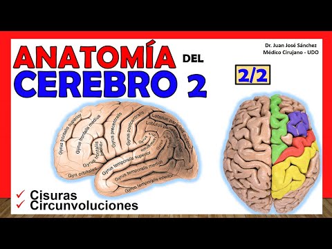
Hello, how are you? Welcome to a new anatomical video on this channel easy anatomy by Juan Jose Sánchez, today I bring you a video about the anatomy of the brain 2, the second installment of this topic that we can also call the anatomy of the brain because precisely with this we begin It is important for you to understand this brain anatomy video 2 in which we will talk about the fissures or also called the sulci, we are also going to talk about the gyri also called convolutions, it is important that you master the faces of the brain that we talked about in the video of brain 1. So I remind you that in order for you to have access to all my slides and to be able to download and use them, I invite you to go to the Pattern link that I leave in the description, then very good without further ado, let's start talking of this brain anatomy 2; The first thing we have to know in this class is that we are going to divide the different faces of the brain for the easiest study of these gyres and these grooves.
In the previous video we talked about how the brain had an external face, also called the face. super external or super lateral, it had a second face that was a medial or internal face and a third face that was the downward one that was the inferior face, so we will specifically start talking about the turns and the fissures that are found on that face external, later we will talk about the internal face to finally finish the video talking about the grooves and turns that are on the lower face, the first thing we have to know roughly when we see a brain from the side, this pole that you see here which corresponds to the temporal pole, he will always look anteriorly, so seeing this point here this tells me that this is the anterior pore, also called the frontal pole of the brain with the brain specifically and this would be the posterior pole. What are we doing?
to find on this external face? Well, we will find a group of fissures that we are going to call intelobular fissures and with it we are going to start the class. Why are they called interlobular fissures?
Because it is these fissures that separate this brain into globules, they are the big ones, let's say, because later we are going to see that each of those lobes is going to be subdivided by several smaller fissures, but these interlobular fissures are the ones that separate me from the globules. four large lobes or well, we are going to see that there are really five that make up the diencephalon, first we are going to see the large lateral fissure also called the Sylvian fissure, that is the eponym that we are going to give it, we are going to see that these fissures It is only seen on the external face but when we see the lower face we are going to see that it is also seen on the lower face of the brain, notice here, here we are seeing a face, an inferior vision that is, we are seeing the lower face, this What we are seeing here is the temporal pole that I was naming you just now, and notice how at the level of the substance or previous perforated space it is said that on the lower face at this level that lateral fissure begins, at this level this fissure separates the lower face, it separates me into two parts, towards the front we have the frontal lobe and towards the back we would have the time lobe the famous temporal lobe, then this fissure starting from this previous perforated space goes outwards, notice how it delimits the two of me lobes and then continues its oblique path upwards and backwards , it is oblique upwards and backwards, this fissure is quite deep, it is said to measure 8 to 9 centimeters in length, it is so deep that we will see later that practically It is covering a large lobe that is hidden in the depths of it, we are going to say roughly that these lateral or sylvian fissures are going to have an extension towards the anterior that invades frontally from the fissure, but see how this extension anterior or anterior branch invades that frontal lobe and we will have an ascending branch that also invades part of that frontal lobe, some books describe a third branch that is the posterior branch, but it is an inconstant branch that we will not always see, this branch before sorry, later It would also be in relation to or invading the frontal lobe, so this portion that you see here on the external face would correspond to the upper portion of the lateral fissure, because the lower portion is the one we saw just now on the lower face, it is important to know that This lateral fissure would be separating my frontal lobe in front of it and behind it the famous temporal lobe that we saw in the first installment of this anatomy of the brain. Notice here how it practically forms opercula in one, like lids.
these two lips, this would be the lid or the frontal lip and this would be the temporal lip, so if I grabbed with the separators and opened through those lateral or sylvian fissures I could find the famous lobe of the insula, which we will study In the final part of this video very well, seen then with this other image we move on to the next internal lobular fissure, notice that the lateral one divided two lobes, let's name them, we are now going to move on to another fissure that also separates my lobe, they are the so-called gross fissures of the brain and we are going to talk precisely about the central fissure, also called Roland's fissure, see that they have many eponyms, so we are going to describe the anatomy of this central fissure but first don't get away from the video [ Music] it is important that you subscribe to the channel below where you see that it says subscribe, click on it and you will automatically be subscribed to the more than 220 videos in it. So we were with this central fissure also called the Roland fissure, notice that this fissure is a little more vertical, it is actually somewhat perpendicular to the cilia fissure and lateral fissure and it heads upwards ending at the upper edge of that hemisphere of each of the hemispheres, in fact, invades, as you can see here, the medial aspect and we are going to see that it specifically invades this lobule called the paracentral lobule, which we will study a little later, approximately the size or length of this central fissure or Rolando is 9 to 11 centimeters, okay? We are also going to see that it is described as having three curvatures, that is, we can see that it is not completely straight, but rather that first it makes a convex curvature forward, then a concave curvature forward, then another convex curvature forward, here we can go into great detail.
Better as it is one convex towards, in front another convex forward and this one that would be concave forward, that is characteristic of this Rolando fissure. Why is the Rolando fissure important? because that is the anatomical point where I separate backwards, remember that this is the parietal lobe and forwards the frontal lobe, so see that it has a great separation, from an anatomical point of view it is the one that separates, from fact that separates the motor part that is responsible for the movement of the brain, such as that frontal cortex, from the sensitive part that is responsible for a little more, the cortex, finally as the internal ocular fissure, we will talk about the parieto-occipital line that some books They call it the parieto-occipital suture, whatever passes from this parieto-occipital line, which in fact is called the external parieto-occipital line, is the full name, even though I didn't put it here, or the external parieto-occipital fissure.
Sometimes it is not so marked and since it is not so marked many times it will happen to us that we are going to see an image like this and we are not going to see it as well as we see the two figures that I named at the beginning, then this parieto line- occipital is a line that we are going to draw from two points, from two two minor fissures in order to delimit the occipital lobe and separate it from the rest. Notice, this parieto-occipital line begins at the beginning of the parieto-occipital sulcus, which is, let's say, a depth that You see here and it ends - be careful, this occipital parietal groove is also seen on the medial or internal surface - if I turn this hemisphere I will see it there and we will see that the same parietal line is also formed there - occipital or whatever you want to call it parieto-occipital fissure but we are no longer going to give it the external name but since it is the middle face we are going to give it the internal name and We are going to see that the point of the antero-inferior edge, sorry, the lateral inferior one is this little groove that is half seen here, so you can see that it is not so marked, that it is the famous pre-occipital notch, then you will see how we draw a line that goes from that notch up to that parieto-occipital sulcus and that is going to separate the occipital lobe that is in green from two things, it separates it in the lower part of the temporal lobe and in the upper part of the parietal lobe; Finally we are going to draw an imaginary line from the end of that lateral or sylvian fissure until this line is perpendicular to this parieto-occipital one in order to delimit the parietal lobe from the temporal lobe, however this is not a fissure, but rather a imaginary line that we are going to draw. Now, what turns do we find at the level of this external face?
Well, let's separate it, so first we talk about the frontal lobe. I remember that the frontal lobe is delimited in front of the central sulcus or fissure and above and in front of the lateral or sylvian sulcus that we are going to find. Notice, first we find two large sulcus, we first talk about the superior frontal sulcus, which is a sulcus that runs more or less parallel to the same axis or the same path that the upper edge of the cerebral hemisphere carries, it is called superior frontal sulcus or superior frontal fissure and we are going to see a lower part which is called the inferior frontal sulcus or fissure, then they are the two large sulcus that separate me from this frontal lobe and we cannot call these sulcus interlobular sulcus, why can't we call them interlobular sulcus?
because in theory I am no longer dividing any lobe, simply now I am making a subdivision of one of the lobes specifically to put it in a more understandable way for you. We are going to see that in the final part of both grooves, I was talking about the upper and lower frontal, they are divided into an ascending and descending branch and at the confluence of these ascending and descending branches, a third groove is formed that we are going to see in relation to This frontal lobe, which is the precentral sulcus, okay? We call it pre- rolancico groove, why pre-rolancico?
because the central groove, which is this one in black, was called the Roland groove, you remember, or the Roland fissure. Well then, they call those grooves gyri, the gyrus are the same as the gyri, let's see then that the gyrus that remains between the upper edge of the hemisphere of the superior frontal sulcus is the superior frontal gyrus, in some books you will see it with the name of first frontal gyrus or first frontal gyrus, then we are going to see this one that is in yellow which would be the middle frontal gyrus this middle frontal gyrus or second frontal gyrus or 2nd frontal gyrus is the one found between the superior frontal sulcus and the inferior frontal sulcus, finally we are going to look at the inferior frontal gyrus, this inferior frontal gyrus would then be delimited between the lateral or sylvian fissure and the inferior frontal sulcus, here I put them in red, it is important to know that in relation to These three frontal gyres we describe three portions, an anterior portion and the frontal gyrus, a middle portion and a posterior portion and this applies to the three frontal gyres, anterior portion, middle portion, posterior portion, to make it a little more detailed Specifically with this lower frontal turn we are going to see that in it we call two large portions a more or less triangular portion, okay? which is this one that I am pointing out to you and a portion posterior to it that is called the operculum portion, so the triangular portion and the opecular portion are the two large portions of this inferior frontal gyrus.
Very well, then we continue talking about the fourth and last gyrus that is in relation to this frontal lobe, which is then the pre-central gyrus, this pre-central gyrus also called the pre-rolling gyrus and with the most important from the point of view of the motor skills of the human body. At some point I plan to make a video of the Brodmann areas and you will see that it is a very important area that is located in this precentral gyrus, notice that this precentral gyrus is parallel to a gyrus that is behind the fissure central so if this turn is called pre central, at once I'll tell you the one behind it is called post central and why am I telling you this? Because there are two places where the pre-central gyrus joins or communicates with the post-central gyrus, the first is in this lower part, yes?
We are going to call it the inferior fronto-parietal operculum, although there is a frontal and parietal fronto because this already corresponds to the parietal lobe, we also call it the inferior fronto-parietal passage fold , it is the same one that I am pointing out here, we see in the middle face, again I have to get ahead of this, let's see that what you see here in the middle is the central gyrus, the central sulcus, sorry central sulcus or rolando so more or less what we see here is the continuation of a sulcus called post central, and this one that we see here in the continuation of the present sulcus seems central, which means that this is the extension towards the medial face of the precentral gyrus and that this is the extension towards the medial face of the postal order. Notice that both turns are also joined this time in the upper part, he spoke of the present, the process, the central post and this is then called the superior frontoparietal percus or also superior frontoparietal step fold, do not forget that the folds of step are the places where turns are made up where different turns join, very well now we are going to talk about the occipital lobe, which remember that it was the one behind this parieto-occipital sulcus, this occipital lobe is actually quite complicated, because some books talk about some grooves but it turns out that in the majority, they talk about how those grooves are very few marked and that is why it is very difficult not to mix them and give them a name, however, where the latarjet converges, which is a little more specialized to talk about of the occipital lobe, first talk about a transverse occipital sulcus ok, that this transverse occipital sulcus, let's see that next to this sulcus called sub parietal sulcus that we achieve right now when we talk about the parietal lobe. What I want you to know is that this transverse occipital sulcus actually crosses the future structure, no forgiveness, temporo-occipital fissure, the one that is supposed to live here crosses the transverse occipital sulcus, then we would have a semilunar sulcus that we can see It has a concavity towards the back and then the calcarine sulcus or the beginning of the fissure or sulcus or calcarine sulcus, those are the three great structures from the sulcus point of view that we can find in the occipital lobe, it is quite difficult to separate gyri or some gyrus in this occipital lobe, which is why it is almost always simply called the occipital lobe and that's it, at least this external side is like that.
So now we are going to talk about the temporal lobe, remember that the temporal lobe was the one that was below and behind the lateral fissure and in this case, in front of the suture, I am going to continue with the suture of the parietal temporal fissure Sorry, occipital temporo which was the one that was more or less at this level, separating the occipital lobe from the temporal lobe in front, look here, we are going to find two large grooves, one is the superior temporal sulcus and the other is the inferior temporal sulcus; This upper temporal sulcus is more or less parallel, which follows a much less tortuous path than the lower one, and which is more or less parallel to the lateral fissure, from the front where the temporal pole is until it practically ends. the temporal lobe and joins the parietal. We are going to see that the inferior temporal sulcus, for its part, is a little more, let's say, tortuous in its path and is parallel to the previous one so that the two temporal sulcus are parallel to the lateral or sylvian fissure, so these temporal sulcus suit me To delimit three large gyri, which are the temporal gyri or gyri, first we would have the first temporal gyrus or first temporal gyrus, which is also called the superior temporal gyrus.
This superior temporal gyrus would be between the lateral sylvian fissure and the superior temporal sulcus, ok? It continues upward with the inferior parietal lobe that we are going to see it now; Then we have the middle temporal gyrus, which is also called the second temporal gyrus or second temporal gyrus, this will be found between the superior temporal sulcus and the inferior temporal sulcus to finally find the third temporal gyrus, which is the inferior temporal gyrus, which is The gyrus surpasses the external face of this, by the way, and we are also going to see it in a part of it on the lower face. This is the famous inferior temporal gyrus.
It is important to know that these gyri of the middle and inferior temporal gyrus also have communication with the inferior parietal lobe, we are going to see right now that they communicate with specific portions, well we are going to see it right below, when we talk about the parietal lobe remember that the parietal lobe is quite difficult to separate anatomically from the occipital there is no anatomical point as such but rather this parieto-occipital fissure, which is almost always imaginary, is very difficult to follow. We are going to see that this parietal lobe is crossed by a fissure that runs from anterior to posterior more or less parallel to the axis of the upper edge of the hemisphere. This is the intra parietal sulcus or they also call it sub parietal sulcus, it is going to separate the parietal lobe from a superior parietal lobe and in an inferior parietal lobe, remember that the anterior limit of the parietal lobe is the central fissure or rolandiac fissure, which is what separates it of the frontal lobe.
Very well, we are also going to see that there is, let's say, a sulcus that is also in relation to this intra-parietal sulcus, which is the post-central sulcus. This post-central sulcus originates at the level of the sylvian fissure and rises to the upper edge of the cerebral hemisphere and invades to the internal face as we had talked about previously, okay? very good so what twists are we going to find here?
As I tell you, the superior parietal gyrus is sometimes called simply the superior parietal lobe because it is quite grotesque as such and we are going to have the inferior parietal gyrus below it. This inferior parietal gyrus has two very important portions, one of which is the supra gyrus. marginal gyrus, which is the gyrus that unites today also the superior temporal gyrus with the inferior parietal lobe and below and behind it we would have the angular gyrus, which is the gyrus that is responsible for uniting the inferior parietal lobe with the middle temporal lobe, eye to both.
supra marginal and angular gyrus are part of the parietal gyrus or the inferior parietal lobe, which I prefer to call it the lobe, because this really does not look like a gyrus like the others, it simply looks like a brain mass and that's it, and we would not have the post central lobe, which is the lobe that is behind the central sulcus, remember that this post-central gyrus joined the pre-central gyrus through the inferior fronto-parietal and superior fronto-parietal passage folds, which was this one. By the way, we took advantage of this image to show them very well, we already talked about the external face a little extensively, now we are going to talk about the internal face of the structures, videos that we find on this internal face or medial face, first we will talk about the interlobular fissures, which separate me from the structures at this level. Let's see first the cingulate sulcus, because sorry, the marginal callous fissure, this marginal callous fissure is made up of two portions, first the cingulate sulcus, which is the same cingulate circus or they also call it singular circus, with c eye, which is one that More or less it goes parallel and the turn of the cingulate which is this one that we are going to see right now ok, and that goes more or less in relation to the corpus callosum, see how more or less the path of the corpus callosum contours it and then when it does an upward curvature becomes a marginal sulcus, be careful, this marginal sulcus is practically the continuation of the central post sulcus, it is practically the continuation, if you follow the continuity of the video this is the central sulcus, and this would then be the post central and there is the pre central; then it would be said that the central post continues as a marginal sulcus, but here it is already part of the marginal callosal fissure.
Then we're going to look at Brock's infra-parietal sulcus , this brock's infra-parietal sulcus, some portion of the marginal callosal fissure that invades practically to the correspondence of the parietal lobe on the internal face, very well we are going to see towards the posterior part of the calcarine fissure, this calcarine fissure has an anterior portion, here we see it looking for the corpus callosum ok, specifically the splenium of the body callosus and a posterior portion, which is what we generally see almost always with the name calcarine fissure and we are going to see the internal parieto-occipital fissure, which is the internal view of the external parieto-occipital fissure that we saw a while ago, see which begins in the same place and ends at the confluence of the calcarine fissure specifically, very well what twists are we going to find here, well look here he marked the marginal callosal fissure, here he marked the internal parieto-occipital fissure, and here the calcarine ; First we are going to find the internal frontal gyrus, which is the internal frontal gyrus, also called the medial frontal gyrus, simply, it is the same superior frontal gyrus but when we see it from the internal side , we give it the name internal frontal gyrus, but it is the same superior frontal gyrus seen on the middle face, important that this internal frontal gyrus is very close to the cingulum gyrus, very close to the part of the face of the corpus callosum ok, very good. Then we are going to see the groove to center, this groove to center is nothing more than the continuation of the pre-central sulcus here we are going to call it the central sulcus ok, let's see then that between that sulcus for the central and the marginal sulcus, remember that it was the continuation of the marginal callous sulcus Well , between the two of them there is a lobule called lobules for centering, important because this beautiful one has part of the parietal lobe and has part of the frontal lobe, we call it lobules for centering, very good, this is where I told you that the union of the precentral gyrus with the postcentral gyrus in the superior frontoparietal operculum or superior frontoparietal passage fold, as I show you here innervate, very good, then we have the symbol gyrus, this gyrus of the good cingulum goes around practically the corpus callosum ok It is very important that there is a separation. Do not believe that this part of the brain is joined to the body, but that there is a groove there that separates them, which is precisely called the groove of the corpus callosum.
Very well, let's see then a region. posterior triangular which is a region found on the medial face of the occipital lobe, which is the famous wedge and a region called quadrilateral lobe or pre wedge, because a more or less square shape that is found practically corresponding to the medial projection, or It is the internal face of the parietal lobe, it is the global extension of that lobe, very well. Finally, to finish with the telencephalic faces, we are going to talk about the lower face , which is much less complex and well, finally, we talk about the hidden lobe, which is the lobe of the insula very well, which is the interlobar fissures we are going to find here, We practically only found the lateral fissure of Silvio, the lower portion of this fissure that we already talked about at the beginning of the video.
This will separate the lower face into the large lobes: one anterior, which corresponds to the frontal lobe, and in turn to the anterior cranial fossa, which is the orbital lobe, and one posterior, which corresponds to the temporal and occipital lobes, that is, the middle cranial fossa and the tentorium cerebellum, notice, we are going to start talking about the anterior portion, which is the orbital lobe, what you see here in green, we are going to see a very medial groove that is the famous olfactory groove, it does not receive another name but the olfactory groove and we are going to see that lateral to it, a groove in the shape of an h, that is, the letter h, called the h-shaped cruciform groove, Latarjet argued in the 1978 version that it should not be called cruciform because It is not really in the shape of a cross but that is how we find it many times in atlases, this cruciform groove delimits several turns, first the internal olfactory groove, which is this one that was indicated in green, then lateral to it between the cruciform groove and the olfactory groove. we would have here in purple, the internal orbital gyrus, then the external or lateral orbital gyrus, then an orbital gyrus anterior and an arbitrary posterior turn, in easy to name if first we limit the sulci, very good, now we move on to talk about the parieto-occipital lobe which was the most posterior portion of let's say this inferior face, first we have the external temporo-occipital sulcus, which sometimes they simply call it the temporo-occipital sulcus and then the internal temporo-occipital sulcus, which sometimes they call it the collateral sulcus but I think it is easier if we use this nomenclature of internal and external, that sulcus that runs from anterior to posterior on that lower face. It is going to divide into two large gyrus, first the external temporo-occipital gyrus and then the internal temporo-occipital gyrus sometimes the internal temporo-occipital gyrus, sometimes they call it hippocampal gyrus with hippocampal gyrus but really we are going to see that the gyrus of the hippocampus is a part, it is a part of this external temporo-occipital gyrus, sorry, internal, it is only a part of it, very well, let's see then what this internal temporo-occipital gyrus is divided into to a more posterior portion, called the lingual lobe and one a little more anterior called the hippocampal gyrus, hippocampal gyrus and then this hook that makes this hippocampal gyrus, which remember that it is the most anterior part of the temporo-occipital gyrus, that hook is called the ungus or hook of the hippocampus, actually the name that the theory says is the ungus of the hippocampus ok, let's see here that there is also a portion that is the isthmus of the gyrus to the symbol that is that, that is the union of the most final part of the gyrus of the symbol very close to the splenium of the corpus callosum, when it joins this very important internal temporo-occipital gyrus, see it here, in any case this of the gyrus of the symbol and see that there is a communication with the internal temporo-occipital gyrus, This union that is here, this step, this step that you see here is what is called the isthmus of the turn of the sign.
Very important that you know that, very well let's see then to finish the video that was a little long but it is really very fascinating and the information is quite long, we are going to open this lateral fissure or silvio so that in order to find the insula And the famous lobe of the insula, that is why I told you that there are actually five globules in the brain, this lobe of the insula, it is said, has a more or less triangular shape, it has delimited edges that we are going to divide in an anterior sulcus, a superior sulcus and an inferior postero sulcus; postero inferior, superior and anterior; That's what makes me the ridge of the insula, that's what separates me from the lobe of the insula and we're going to see that diagonally from the superior posterus to the inferior antero, it has a groove that separates me from that achievement to the insula, which It is the central sulcus of the insula, it is important to know that this central sulcus of the insula separates the insula into an anterior lobe and a posterior lobe which is the one here in lilac, then the anterior lobe in yellow and the posterior lobe in lilac and to complicate it even more we are going to see that the anterior lobe through two grooves is separated into three turns or convolutions, one anterior, one middle and one posterior; The tip where these three turns of the anterior lobule converge is what is called the pole of the insula, the tip to pole of the insula and then the posterior lobule; It only has a groove that separates it into a gyrus, an anterior gyrus, and a posterior gyrus or gyrus. Notice here in this section how we can see the insula that is practically inside, deep inside each of the cerebral hemispheres. Something also important that you have to know is the front wall of this gray substance, which is buried in the nuclei.
The progress of this substance is, let's say, gray tissue that is found in that front wall between two structures of white substance, one is the external capsule that you see is white and one is the other in the extreme capsule that you see is also white, so this substance which is located at the base of the insula, what you see is the insula, is specifically the front wall and that is why they are named, named because of the very important anatomical relationship it has with the base of the insula, This was then the entire video of Brain 2, I hope you liked it, don't forget to subscribe and like the video if you liked it.
Related Videos

15:29
🥇Anatomía del CUERPO CALLOSO. ¡Explicació...
Anatomía Fácil por Juan José Sánchez
64,019 views

19:21
🥇 ANATOMY OF THE BRAIN 1/2. (Telencephalo...
Anatomía Fácil por Juan José Sánchez
367,352 views

17:44
🥇 SISTEMA NERVIOSO PERIFÉRICO (S.N.P) - G...
Anatomía Fácil por Juan José Sánchez
184,268 views

1:36:02
V. Completa. Cómo funciona nuestro cerebro...
Aprendemos Juntos 2030
3,625,667 views

Classical Piano & Fireplace 24/7 - Mozart,...
Odd Eagle

19:45
Así es como recuerdo TODO lo que aprendo e...
Tutorías Medicina Interna
954,503 views

22:09
Cerebral Cortex (Function, Covering, Lobes...
Taim Talks Med
212,011 views

12:26
🥇 Anatomy of the DIENCEPHALO. (Generaliti...
Anatomía Fácil por Juan José Sánchez
139,879 views

12:02
How Does Music Affect Your Brain? | Tech E...
WIRED
741,337 views

30:54
🥇 COXAL BONE - ILIACO BONE, Anatomy. Easy...
Anatomía Fácil por Juan José Sánchez
485,615 views
![Lobes of the Brain: Cerebrum Anatomy and Function [Cerebral Cortex]](https://img.youtube.com/vi/tZFW-waIpQg/mqdefault.jpg)
19:45
Lobes of the Brain: Cerebrum Anatomy and F...
EZmed
738,358 views

16:46
Irrigación cerebral
Juanse Rodríguez MD
261,126 views

11:54:57
Beautiful Relaxing Peaceful Music, Calm Mu...
Tim Janis

53:33
Your Brain: Who's in Control? | Full Docum...
NOVA PBS Official
4,652,012 views

41:45
Neuroanatomía seccional básica (TAC y RM) ...
Radiología e Imagen para Estudiantes
245,248 views

12:41
🥇 Anatomía del FÓRNIX (Trígono Cerebral)....
Anatomía Fácil por Juan José Sánchez
42,532 views

13:56
The Brain
Bozeman Science
5,918,342 views

4:40
Anatomía del Cerebro en 3D, Animación. Ali...
Alila Medical Media en Español
931,091 views

25:56
Ventricles of the Brain: Anatomy and Cereb...
EZmed
189,125 views