Oral Vestibule (Lips, Cheeks, Teeth, Gums) - Oral Cavity Anatomy
154.35k views2066 WordsCopy TextShare

Taim Talks Med
Content:
0:00 Introduction
1:57 External Structures of the Mouth
2:49 Division of the Oral Cavity
3:...
Video Transcript:
What’s up, Meditay here, Let’s talk about the anatomy of the oral cavity. Now since this is my first video of the digestive system, I wanna spend a quick minute giving you a little overview of the whole digestive system. And to do that, we’ll use this chocolate cheesecake to highlight al the structures it’s gonna go through within your digestive system.
The reason why I chose a cheesecake is because it most probably starts you salivating because you probably wanna eat it. That implies that the Oral cavity is the first part of the digestive system. After the oral cavity is the Pharynx.
Then when you swallow the food, it’s going to go through the esophagus, and then all the way down to your stomach. After it’s been processed by the hydrochloric acid in the stomach, it’s then going to enter the small intestine, which consists of the duodenum, then the jejunum, and then the ileum. And after the Ileum, it’s going to enter the Large Intestine, which consists of the caecum and the colon, and then the rectum.
And by the time it gets to here, this is how the cheesecake’s gonna look like. And the fuller the rectum gets, the higher you feel the urge to poop. So those are all the structures of the digestive system.
But we also have a system of accessory structures which help us digest the food we eat. Those include the Teeth, Tongue and Salivary glands. And then in the abdominal cavity, there’s the Liver, the Gallbladder and the Pancreas.
Our goal is to go through the detailed anatomy of all of these structures, one by one. And we’ll start with the oral cavity. So in this video, we’re first going to talk about the main external structures of the mouth.
Then we’re gonna go through the division of the oral cavity, basically how we divide the oral cavity. After that, we’re gonna talk about the structures associated with the borders of the oral vestibule, which include the anatomy of the Lips, Cheeks and the Teeth. Then in the next video, we’ll focus on the other part of the oral cavity, which is the oral cavity proper.
So here you see the external mouth. You probably didn’t know this, but here’s the upper lip, and here’s the lower lip. They both meet at the lateral ends as the Oral angle, or the labial commissure.
Above the lips, you're gonna have two grooves on either side, as you see here, called the Nasolabial Sulcus. And then in the middle, there's going to be a depression called the Philtrum. And then under the lips, there's going to be another Sulcus, called the Mentolabial Sulcus, or mentolabial Fold.
Now, remember the muscle that's located inside of your lips? The Orbicularis Oris Muscle? This muscle is going to help regulate the opening of your Mouth, And that opening is called the Oral fissure, or in Latin Rima Oris, and that my friends are going to be the opening of our digestive pathway.
Now. We generally divide the oral cavity into two parts. There's the Oral Vestibule here in green, and the Oral Cavity Proper, here in blue.
And the dividing line between these two parts are gonna be the teeth and the gums. Alright. So again, In this video, we're mainly gonna focus on the Oral vestibule.
And we’re gonna do that by going through the structures associated with the external and the internal borders. The external borders of the Oral vestibule are going to be the Lips and the cheeks. And the internal borders of the oral vestibule are the teeth and the gums.
Awesome. So let’s go through the anatomy of these, starting with the Lips. Now, the most basic things you need to know about the anatomy of the lips is that the inner and outer sides of the lips are covered by different types of epithelium; there is a transitional zone between the two sides The upper lip is attached to the gums through the Frenulum of the upper lip.
And the lower lip is attached to the gums through the Frenulum of the lower lip. And within the mucosa of the lips, you’ll find minor salivary glands called labial glands, which continuously produces saliva that lubricates the inner side of the lips. And keep in mind the structures we went through earlier; they’re also associated with the upper and lower lips.
Awesome. Now let’s talk about the cheeks, or the Buccae. The shape of the cheek is formed by the buccinator muscle, which is this one.
On top of the buccinator muscle, there’s a fascia called the buccopharyngeal fascia, which remember covers the buccinator, then goes backwards behind the pharynx. On top of the Buccopharyngeal fascia, you’ll find the buccal fat pad (Bichat’s fat pad). The buccal fat pas, together with the subcutaneous connective tissue gives the cheek its definitive appearance.
Now externally to the fat pad, you’ll find all the layers of the skin. And on the inner side of the muscle, you’ll find the mucous membrane, or tunica mucosa. Now the tunica mucosa of the cheeks, at around this region, you’ll find the opening of the parotid duct, which is the duct of the parotid gland as you see here.
And around the opening of the parotid duct, you’ll find the Papilla of the parotid duct. Which is just gonna be a small elevation around where the parotid duct opens into the oral vestibule. And here just to help you visualize it.
Imaging for a second that this hole is the opening of the parotid duct. And this is how the papilla of the oral vestibule looks like. Just some elevations around the opening.
So that was everything I had for the anatomy of the cheeks. Now let’s cover the Inner surface of the oral vestibule and start with the anatomy of the teeth. So, let’s take this tooth right here, and pull it out.
And then slice the tooth in half in order to see the inside as well. The tooth actually have a very straight forward anatomical structures despite its comprehensive development. It’s usually divided into three regions.
First we have the Crown of the tooth, which is the visible part of the tooth. Then we have the Root of the tooth which is imbedded in the dental alveolus. And between these two parts is a neck, which is the transition between the crown and the root.
The neck is covered by the gums as you see here. Now. The root of the tooth is fixed inside the dental alveolus, right`?
And this fixation is supported by something we call periodontium, which forms the so called gomphosis, which is a vert strong fibrous connection between the root of the tooth and the periosteum on the inner surface of the dental alveolus. The gomphosis is a type of joint we call the dentoalveolar joint. Within the actual tooth, there’s a cavity.
And within the cavity, you’ll find the dental pulp, which is a type of loose connective tissue that is rich in neurovascular structures. The pulp cavity will continue downwards as the root canal, and then at the very bottom of the root, there’s an opening called the apical foramen, which is where blood vessels and nerves from enter the tooth. Around the pulp cavity, you’ll find the Dentine, which makes up the basic substance of the tooth.
And at the crown region, the dentine is covered by the enamel, which is a very mineralized tissue. And in the region of the neck and the root, you’ll find the Cementum lying just superficial to the enamel, which is a calcified structure around the enamel. And here’s a little more detailed view of the layers around the root of the tooth.
First you have the Dentin. Then the Cement around the Dentin. And then around the cementum, you’ll find the periodontal ligament.
So that’s the general anatomy of the tooth. Now. Throughout out lives, we have two different sets of teeth.
The first set of teeth are called milk teeth, and there’re 20 milk teeth usually. They begin to show up at around 6 months of life, and then by the time we’re 24 months old approximately, we should have a full set of milk teeth. As we start to grow.
The milk teeth are gradually starting to fall off between the age of 6-12 years, and they start to get replaced by the permanent teeth. The permanent teeth consist of 32 teeth, and the last set of teeth which appear at around the age of 17-24 years are called the wisdom teeth. They’re called wisdom teeth because they appear later, after all other teeth have appeared.
So milk teeth and permanent teeth. Now. The arrangement of the teeth are categorized in a specific way.
I learned recently that the way we divide the teeth in Europe might differ from the way the teeth are divided in the US, so keep in mind that this might differ depending on where your sources are from. I study in Europe so I’ll go with that. First, we divide the teeth into four equal quadrants.
Starting from the midline. The first two teeth from the midline are called the incisor teeth. And they get the numbers 1 and 2.
So you start counting from 1 at the midline and then 2 and then 3 and so on. So when I say we have two incisor teeth, I mean we have 8 incisor teeth, right? Since you count 2 from midline on both the upper and lower row.
The Incisor teeth consist of only one root and usually only one root canal. So the 3rd tooth is called Canine tooth. Which also contain one root.
So we have 4 canine teeth. Then number 4 and 5 are called the Premolar teeth, and number 6, 7 and 8 are called Molar teeth. The last one being the wisdom tooth.
And if you count 8 teeth on each quadrant. So 8 times 4 equals 32, that means that we’re looking at the permanent teeth now. Since remember the permanent teeth consists of 32 teeth.
The milk teeth differ a little bit, but they’re divided in the same way. They have 2 incisor teeth. 1 canine teeth and 2 molar teeth.
So they don’t have any premolar teeth. And that is the main difference between the milk teeth and the permanent teeth. And here you see that the incisors and canine teeth usually have one root, but the premolar and molar teeth might have more roots as you see here.
Now. Let’s talk about the gums, or the gingiva! From the anatomical point of view, the gum is a mucous membrane covering the alveolar processes of the maxilla and Mandible.
And they form a place for the teeth to be rooted in as you see here. Now, the gums are mainly composed of dense fibrous tissue, but the actual consistency of the gums differ depending on which region of the gums you’re looking at. From the clinical point of view the gum is divided into the alveolar mucous membrane, and the gum proper.
The alveolar mucosa covers the alveolar processes at the level of the root of the tooth, as you see here in blue. This region contains a lot of submucosal connective tissue and is therefore mobile. So, it moves if you touch it.
The gum proper is the mucosa located at the same level of the neck and crown of the tooth, as you see here in yellow. The mucosa itself doesn’t have a lot of connective tissue here at the region of the gum proper, because it’s attached to the periosteum underneath. That’s why if you try to move the gums at around the neck of the tooth, you’ll see that it’s immobile.
The gums here don’t move at all. Anatomically, you can divide the gums into certain regions. The gingival papillae are the projections you see between the necks of the teeth.
The gingival margin is the elevated margin of the gum around the tooth And the gingival sulcus is the groove between the margin of the gum and the tooth So that was all I had for the anatomy of the Oral vestibule. In the next video, we’ll cover the anatomy of the Oral cavity Proper.
Related Videos
16:14
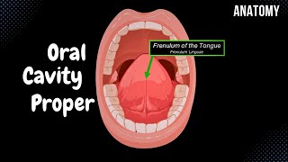
Oral Cavity Proper (Palate & Tongue) - Ora...
Taim Talks Med
243,224 views

26:20
Gastrointestinal | Development & Embryolog...
Ninja Nerd
1,075,785 views

14:37
The Science of Building Your Pecs: Best Ex...
Institute of Human Anatomy
137,968 views

Relaxing Music to Relieve Stress, Anxiety ...
Soothing Soul
12:16
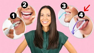
The Perfect Oral Health Care Routine (3 ea...
Teeth Talk Girl
443,520 views

16:30
Blood and nerve supply of the oral cavity
Osmosis from Elsevier
94,987 views
18:19
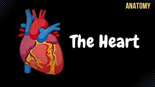
Anatomy of the Heart - External & Internal...
Taim Talks Med
673,422 views

13:42
Dental Anatomy | Maxillary Central Incisor...
Mental Dental
85,465 views

44:04
Gastrointestinal | Salivation: Parotid, Su...
Ninja Nerd
526,038 views

24:42
Peritoneum tutorial
The Noted Anatomist
1,891,969 views
18:12
We FINALLY Found a Way To Starve Cancer
Dr Ben Miles
389,273 views

14:59
Oral cavity anatomy
Sam Webster
88,279 views

16:59
Peritoneum (Parts, Lesser & Greater Omentu...
Taim Talks Med
562,465 views

40 Hz Binaural Beats: 100% Brain Activatio...
Good Vibes - Binaural Beats
15:19
Dental Anatomy | Maxillary First Molar | I...
Mental Dental
46,066 views
21:36
Large Intestine Anatomy (Parts, Topography...
Taim Talks Med
139,615 views

18:18
Cranial Nerve BASICS - The 12 cranial nerv...
ICU Advantage
1,680,282 views

17:05
Basic Eye Anatomy and Physiology
Dr Matt & Dr Mike
280,909 views

25:08
Introduction to the gastrointestinal tract...
Sam Webster
99,820 views

41:33
Digestive system
The Noted Anatomist
1,024,370 views