Lung Carcinoma (Lung cancer)
446.38k views1628 WordsCopy TextShare
Armando Hasudungan
Support me:
🖼️ Buy PDFs: http://armandoh.org/shop
💵 Patreon: http://www.patreon.com/armando
👕 B...
Video Transcript:
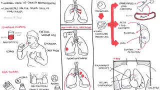
lung cancer is a leading cause of cancer related deaths and it has a poor prognosis in this video we will focus on lung carcinomas so let us look at the different types of lung carcinomas and it can be divided into two broad types small cell car carcinoma and non small cell carcinoma let us focus on small cell carcinoma small cell carcinoma represents about 15% of of these of L carcinomas more than 60% actually present already with um metastasis the prognosis of small lung uh small cell lung carcinoma is poor the tumor tends to grow proximately
close to the hilum and involves neuroendocrine cells in the area because neuroendocrine cells are involved in this type of cancer they undergo mutations which allow them to produce hormones like hormone likee substances that they should not be able to produce and so as a result they release these hormones and it triggers a phenomenon known as the Paran neoplastic syndrome which we'll talk about later on nons small cell carcinomas represents the majority of lung carcinomas 85% non-s small cell car caroma is further divided into three types adenocarcinoma is the most common type of non-s small cell
carcinoma adenocarcinomas make up 38% of lung carcinomas these types of cancers tend to occur in the peripheral lung tissue so away from the hilum and involves glands within the lung squamous cell carcinoma is the other type of non-s small cell carcinoma and makes up about 20% of long carcinoma cases making it the second most prevalent type of L carcinoma these types of cancer tend to occur close to the main broncus and can cause obstruction of the Airways they are called squamous because the epithelial cells that line in the airway become mutated and change from columna
cuboidal to squamous and essentially dysplasia cancer the last type of non-s small cell carcinoma is large cell carcinoma which make up about 5% of L carcinomas so it is the least common large cell carcinoma rapidly grows like the small cell carcinoma and can present in the periphery the peripheral lung tissue or the proximal long tissue so those were the different types of long carcinomas let us look at the signs and symptoms of patients that present with long carcinomas now not everyone presents um like with these uh with with the same signs and symptoms but the
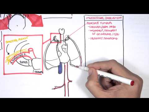
most common signs and symptoms include cough weight loss hemoptisis disnea and chest pain there are many risk factors for lung cancer major ones are smoking radon air pollution arsenic T asbestos nickel as well as this family and genetic f factors now with cancer cuz it is a growth it can cause some problems to surrounding tissues surrounding organs so let us look at some mediastinal involvement of um of lung cancer so let us zoom into the medi mediastinum uh which uh which where we can find the heart the lungs and all these other structures including the
ribs let us zoom into this first rib area and learn a bit anat a bit of anatomy um so here is the sternum and the first rib now going over the first rib are some important structures including the subclavian artery and vein and the brachial plexus the nerves that inovate the Upper Limb why are we talking about these structures well because these structur have to do with the uh with the pathophysiology or the complications associated with lung carcinomas lung cancers see the mediastinal involvement of lung cancer include Panos tumor growth which is growth on the
appical lung surface so on the top which can block part of the brachial plexus depending on how much the brachial plexus is affected it can cause shoulder arm pain weakness and atrophy on the ipsilateral side so on the same side aicle lung tumors can also block a the sympathetic nerve fiber around this area causing U what's known clinically as Hornet syndrome another media styal involvement is plural infusion which causes disa as well as chest pain there can also be heart involvement causing uh pericardial uh infusion another important structure that can be infected is the superior
venina Cava which when blocked can cause the vena cava syndrome forget about the inferior venina Cava here this is a complete mistake ignore that so it's Superior venina Cava so those were the so those were some medial involvement associated with cancer lung cancer growth let us look at the airway involvement now so the Airways of the lungs are the bronchi in the bronchioles right before it terminates at the alvioli well cancer can cause Airway obstruction as it impedes air flow Airway obstruction leads to disia when there is Airway obstruction or irritation this actually sends sensory
information to the brain and triggers the cough reflex that is why in presentation we have disia and cough the patients have disan cough cancer or tumor also stimulates angiogenesis which is blood vessel growth however angiogenesis forms leaky and tortuous vessels which when ruptures Le can lead to what leads to hopsis okay let us zoom into the blood vessels the blood vessels contain your red blood cells platelets and white blood cells right in lung cancer we see some blood involvement mainly anemia which leads to fatigue and disia thrombocytosis in 15% of cases and hypercoagulable disorders lung
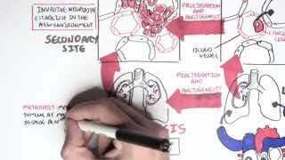
tumors can metastasize and they do when it metastasizes it goes to the heart and then the heart will pump the chuma the the growth the cancer either up to the brain and upper limbs or down to the abdomen area common sites of lung metastases include the brain the liver the adrenal glands and the bone metast metastatic sites are commonly uh asymptomatic now looking back to the different types of lung cancer remember the neuroendocrine cells that begin secreting hormones in the small cell carcinoma um well it leads to the phenomenon called the Paran neoplastic syndrome let
us learn a bit more about what this encompasses so the paraneoplastic syndrome typically occurs in small cell lung cancer as well as squamous cell carcinomas paraneoplastic syndrome are syndromes that occur not related to Invasion obstruction or metastasis of primary tumor and they and the paraneoplastic syndrome include the following ectopic Cushing syndrome where the hormone released by cancer cells stimulate the adrenal glands to produce cortisol we also have antidiuretic like substance which stimulates uh secreted by the Neo Endocrine cells which stimulate the kidneys to retain water these neuroendocrine cells also produce a parathyroid hormone like substance
which stimulates the bone to break down its minerals and release calcium into plasma increasing blood calcium levels resulting in hyper calcemia paraneoplastic syndrome also include uh the hyper pulmonary ASO orthopathy leading to clubbing and periostal proliferation of the tubular bone and lastly inflammatory myopathies um can result from uh lung cancer which leads to the muscle weakness and so that is why we see signs of finger weakness upon examination of patients with lung cancer okay so that was essentially the pathophysiology now lung tumors can be staged we will not look at the staging in this video
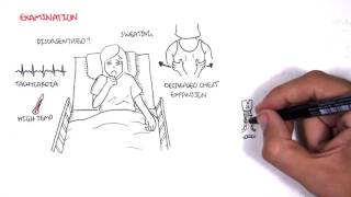
because all I will do is regurgitate what I wrote but I will draw it out uh out quickly and leave you to interpret it yourselves next let us look at some investigations uh we would do if we were suspicious of lung cancer so lung involvement we always do a chest x-ray to rule out other differentials common clinical findings on x-ray for lung cancer include ayum enlargement pulmonary opacity which represents the tumor three rib bone lesions plural fusion and also lung collapse another investigation which is critical for uh for this is c for lung cancer is
CT scan and should be performed early to determine stage and management of the cancer not only CT scans but biopsy are to be performed which include uh the bronchoscopy which is where the prim lung tumor is visualized and Sample is taken using the instrument you can also perform a CT guided fine needle biopsy which is a more reliable way to obtain a histological diagnosis um a needle aspiration this is where a needle is inserted in the lump of the on the lung or lymph node to see for lymph node involvement another form on of Investigation
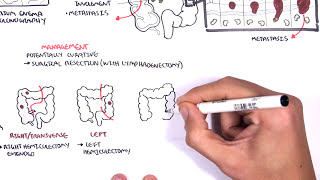
for biopsy is the arthor centesis which is where fluid is collected from the plural cavity and this is used for sampling again uh numbers three four five and six are used for biopsy to Stage the tumor um so that appropriate management can be taken and so management is the next topic we will talk about so these are one surgical treatment which is the most important surgical treatment is for the removal of tumor of the lung tumor cancer for stages one and stages two after surgery or if surgery cannot be performed there is also radiotherapy and
chemotherapy as well as laser therapy and stenting radiotherapy is less effect is less effective than surgery however radiotherapy is used in combination with chemotherapy for stages three chemotherapy increases survival up to one year nausea and vomiting are side effects these side effects are managed best by the 5 ht3 receptor antagonists because these drugs will Target the chemo receptor trigger Zone thus preventing the vomiting nausea Associated symptom laser therapy and stenting can also be done Airway obstruction from the tumor growth causing serious symptoms can be managed using laser treatment and stenting so essentially the obstruction of
the airway you basically remove it so air flow can uh can return as normal I hope you enjoyed this video on lung cancer thank you for watching
Related Videos
8:48
Colorectal Cancer - Overview
Armando Hasudungan
741,775 views
30:19
Lung Cancer/Tumors
Dirty Medicine
162,762 views
13:08
What happens in every stage of lung cancer...
Macmillan Cancer Support
77,399 views

1:35:03
Lung Cancer
Ninja Nerd
133,786 views

11:47
Esophageal Cancer (Year of the Zebra)
Osmosis from Elsevier
79,901 views
16:09
Pleural Effusion (DETAILED) - (pathophysio...
Armando Hasudungan
376,034 views
13:34
LUNG CANCER- Part 1- Epidemiology, Etiopat...
ilovepathology
121,542 views
11:40
Interstitial Lung Disease (ILD) - Classifi...
Armando Hasudungan
212,306 views
![The Cell Cycle (and cancer) [Updated]](https://img.youtube.com/vi/QVCjdNxJreE/mqdefault.jpg)
9:20
The Cell Cycle (and cancer) [Updated]
Amoeba Sisters
5,062,068 views
22:41
S1 Episode 5: Small Cell Lung Cancer: What...
Medscape
495 views
4:58
Lung Cancer - Overview
Armando Hasudungan
194,911 views
16:06
Community Acquired Pneumonia (DETAILED) Ov...
Armando Hasudungan
235,873 views
9:01
How Smoking Kills
Nucleus Medical Media
12,450,497 views
9:02
Cancer - Metastasis
Armando Hasudungan
302,857 views

27:32
Lung Cancer Staging
Radiology Frameworks
21,824 views

28:05
Lungs Cancer Symptoms, Pathology, Treatmen...
MedNerd - Dr. Waqas Fazal
9,177 views

8:25
Cervical Cancer: Osmosis Study Video
Medscape
149,629 views
11:24
Oncogenetics - Mechanism of Cancer (tumor ...
Armando Hasudungan
959,547 views
10:08
Emphysema (chronic obstructive pulmonary d...
Osmosis from Elsevier
1,142,151 views
27:07
Lung Cancer Types
Radiology Frameworks
36,640 views