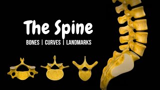
External Spinal Cord (Surface, Segments, Spinal Nerve, Enlargements, Reflex Arch) - Anatomy
175.19k views2622 WordsCopy TextShare

Taim Talks Med
Content:
0:00 Introduction
1:07 Topography of Spinal Cord
2:44 External Surface of Spinal Cord
3:41 ...
Video Transcript:
What’s up. Meditay here. Let’s talk about the anatomy of the Central Nervous System.
In this segment, we will be talking about the external anatomy of the spinal cord. basically, go through everything you need to know in regards to what the spinal cord is and what you’ll find grossly on the spinal cord. Alright, so the Central Nervous System consists of two parts.
The encephalon, and the spinal cord. So in this is video, we’re first going to go through the Topography of the Spinal cord, basically where it is, where it starts and ends. Then we’ll focus on the external surface of the spinal cord, basically going through all the grooves and fissures you see there.
We’re also going to go through the segments of the spinal cord and look at its relationship with the vertebral column. Then we’ll go through the enlargements we see on the spinal cord. After that we’ll look at the anatomy of a spinal nerve, and understand its 4 branches, and then quickly understand the types of reflex arches we can have through the spinal cord.
The internal structures and all the nuclei and tracts will be covered in the next video so that this video doesn’t get too long. Alright, so here we see a posterior view of the vertebral column, if we remove one vertebra and zoom in, you’ll see the spinal cord right here, going through the vertebral canal. So let’s go ahead and take the spinal cord out.
Now the spinal cord is covered by a meningeal layer called Dura Mater. And if we remove the dura mater, you’ll find the Arachnoid mater, and if w remove the arachnoid mater, we’ll see a very thin and delicate connective tissue covering called the Pia mater, and if we remove that, we’ll finally get to the actual spinal cord. These three are what we call Meninges and they cover the whole central nervous system.
We’ll go through these in a separate video. But now, let’s do the topography of the spinal cord. So the spinal cord starts off at the Foramen Magnum, all the way to the L1/L2 vertebra region.
The length of of spinal cord varies a lot, but in general it’s about 40-45cm long. If we now remove the bones, you’ll see that the spinal cord ends by a structure called the Medullary Cone, or Conus Medullaris, from here, a very thin thread goes out called Flium Terminale, which literally translates as the terminal thread because it’s consider as the continuation of the spinal cord. And if we see here, the Filum Terminale continues downwards together with a lot of different nerves supplying the lower part of your body with nerves.
And this area with all the nerves, is called Cauda Equina. So that was it for the topography. Let’s now take a small part of the spinal cord and look at the external surfaces of it.
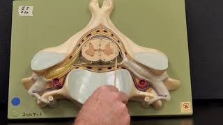
So here is the external surface of the spinal cord. This is the internal surface of the spinal cord which we will talk about in the next video, and these nerves we see here on the sides are the Spinal Nerves. Alright, now for some orientation.
The anterior part is where you’ll find this deep fissure, and the posterior part is more flat, with small bumps. This fissure we see on the anterior side, is called the Anterior Median Fissure. Then posteriorly on the midline, you’ll find a Posterior median sulcus.
On the sides of the spinal cord, you’ll find the Right and Left posterolateral sulci, and the right and left, anterolateral sulci. From where the anterior and posterior root of the spinal nerve are going to go through as you see here, we’ll get back to this later when we talk about the spinal nerve. But now, let’s talk about something called segments.
Alright, so the vertebral column consist of 7 cervical vertebra, 12 thoracic vertebra, 5 lumbar, 5 sacral and this varies, but usually you have 4 coccygeal bones fused together. Adding a total of 33 vertebraes. Now the spinal cord is different in that, it’s divided into 8 cervical segments, not 7, but there’s still 12 thoracic segments, 5 lumbar, 5 sacral, but then only 1 coccygeal segment, which adds up to 31 spinal cord segments.
Now why is the spinal cord divided into segments? Because at each segment, there’ll be one spinal nerve emerging from either side, like you see here. Meaning that picture we saw earlier, with one spinal nerve going out, is one segment.
So we have 31 of those you see here. And if we add the nerves from my 3d anatomy program, you’ll see that it looks like this. Now if we take the vertebral column and the spinal cord, and fuse them together, you’ll see this.
You’ll see the spinal nerves going out from the vertebral column through the Intervertebral openings, or the intervertebral foramina. But if the spinal cord has 31 segments, and the vertebral column has 33 vertebra. And as we know now from the topography, with the spinal cord ending at L1/L2 region.
How are the spinal nerves arranged within the vertebral canal? And to answer that, we need to look at the spinal cord and the vertebra from this perspective. Let’s now fade this picture a little bit, and go through them part by part.
At the beginning, the spinous process of the cranial cervical vertebra, cranial meaning up towards the head, correspond to the same level as the spinal cord segments, and the spinal nerve leave above the first cervical vertebra as you see here. Then as we continue slowly downwards. You’ll see that the spinal nerve starts to bend.
And now, the spinous process of the caudal cervical vertebra, caudal meaning towards the tail, or away from the head, it correspond to one above the corresponding cervical spinal cord segment. And by this I mean Vertebra C7 is at the same height as Spinal segment C8. So you could take Segment +1 at this point.
Because the spinal cord is getting compressed. Then as we continue down, the spinal nerves bend even more and at this point, the spinous process of the cranial thoracic vertebra, correspond to the thoracic spinous segment +2. So you add two numbers to the vertebra you’re looking at, so vertebra T3, is at the level of Spinal segment T5.
And as you slowly continue down, the differences start to be greater. The Caudal thoracic vertebrae start to correspond to the Spinal segment +3… and then as you continue further down. .
the T10-T12 vertebra becomes at the level of L1-L4 spinal cord segment, and then at the Vertebra T12, L1 we’re starting to reach the end of the spinal cord, but not yet, so we call this area the Epiconus, because remember the medullary cone is at the end, epi means above, so above the end of the spinal cord. That’s what epiconus mean. This correspond to L5-S2 spinal cord segment, and then lastly at the L1/L2 region, you have the rest of the spinal cord, from S3 to S5 plus the one coccygeal segment.
This scheme is just to help you visualize how the spinal cord is arranged within the vertebral column. Now. As you look at the spinal cord anteriorly, you’ll notice two distinct enlargements.
One called the cervical enlargement, or intumenencia cervicalis. Which goes from the segment C3 to T2, and a lumbosacral enlargement, or intumenencia lumbosacralis, going from T12 down to the medullary cone. Now why are these significant?
Because at these regions, you have a bundle of nerves called plexuses, supplying the upper limb and the lower limb with nerves. And these nerves have to be large and in a large quantity, in order to innercate all the muscles of the lower limbs and the upper limbs with nerves. So the cervical enlargement forms the brachial plexus for the nerves that goes to both arms, and the lumbosacral enlargement is for the sacral and lumbar plexuses, innervating structures in the pelvis and the legs.
So these are very important. Now since we’re talking so much about the spinal nerves, let’s really understand the anatomy of the spinal nerves. Because once you understand that, the actual internal and external surfaces of the spinal cord becomes more logical.
So if we take a segment of the spinal cord again, you’ll see the internal surface here. We will talk about this in detail in the next video, but the internal surface consist of grey matter, and white matter. And how does this coorelates with the spinal nerve?
Because all the small neurons that go within the spinal nerves, will synapse with nuclei in the grey matter. And I’ll show you how. So first you need to understand where the spinal nerves enter the spinal cord.
The spinal nerve enters the spinal cord through the Right and left Posteriolateral sulci, and the right and left anterolateral sulci. Now lets animate it a little bit and add some structures to make it look a little more realistic. Here we can see the meninges.
So the red that’s closest tot eh spinal cord, is the pia mater, the blue lining is the Arachnoid mater. And between the pia mater and the arachnoid mater is the subarachnoid space, filled with cerebrospinal fluid that provide nutrients to the spinal cord tissue. And then the outermost dense structure is the dura mater.
So these are the meninges. And remember, the deep fissure is anterior, and the more flat surface is posterior. Now here you see the spinal nerve.
The spina nerve is divided into two roots before it enters the spinal cord. It’s divided into the posterior root or the sensory root, and it’s divided into the anterior root, or the motor root. And I’ll mention this again, because I really want you to not forget this.
Posterior root enter through the right and left posterolateral suci, and anterior root enter through the anterolateral sulci. But you’ll notice that on the posterior root, there’s a bulb, a small enlargement called a Spinal ganglion, or sometimes referred to as dorsal root ganglion. You’re gonna have many ganglions in the body, and the reason why ganglions are bubbly, is because they contain many nerve cell bodies as you see here.
So dendritic fibers go from the periphery towards the spinal ganglion, and then the axons of these neurons go into the spinal cord. Now remember from previous video when we went through the different types of neurons. What kind of nerve do you think this is?
These are pseudounipolar neurons, going into he spinal cord, so they take sensory information from anywhere in the body, and then enter the spinal cord so that you can sense what’s happening. So if you blow on your arm, that cold sensation is gonna enter the spinal cord through this neuron. And once it enters the grey matter of the spinal cord, it can either synapse with an interneuron and go further up towards your higher senses so that you can make sense of what’s happening, or it can connect directly to a motor neuron, which go out from the spinal cord, to move a muscle to react in any way.
And there’s gonna be a lot of connections to the motor neuron. Any voluntary movement you wanna do, either comes form the interneurons ro the sensory neurons directly. So a spinal nerve consist of Sensory fibers, Motor fibers, and either sympathetic or parasympathetic nerve fibers.
So in the spinal cord, you’ll find the sensory fibers back here, the motor fibers are in the front here, and the sympathetic and parasympathetic fibers, come from the lateral part fo the spinal cord. And they come from specific areas within the spinal cord. The segments C8 to L2, are responsible for sympathetic nerve response, while the segments S2 to S4 give parasympathetic nerve response.
And here’s a quick way to remember this. S, stands from stress, to remind you that sympathetic neurons are responsible for fight or flight response, or stress response, meaning it increases your heart beat, makes you breath faster, more alert, all of those reacions are gonna come from fibers that leave your spinal cord between the C8 and L2 segments. The P in Parasympathetic stands for Peace, which is rest and digest.
SO you’re chilling, you’re sleeping, your intestines are doing its work to absorb the food, and all of those things, come from the neurons that emerge between S2 and S4 spinal segments. Now back to the spinal nerve. So once the spinal nerve leave the spinal cord.
It branches out into 4 parts. It becomes a ventral branch, which supply skin and muscles of your limbs and the anterior and lateral part of the trunk. And as they do that, they form plexuses.
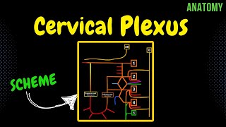
We’ll go through this when we go through the peripheral nervous system. But plexuses are a huge network of neurons that supply regions of your body. So we have a cervical plexus supplying areas associated with your neck and shoulders, the brachial plexus for your arms, the lumbar plexus for your legs and pelvis and the sacral plexus for your pelvis and legs aswell.
So that is the ventral branch, forming these plexuses. The spinal nerve is also going to divide into a dorsal branch for the skin and muscles of the back and neck. Then there’s a white ramus communicans, which relay sympathetic nerves.
And those nerves are involuntary, so they reach out to organs you’re not in control of, like your smooth muscles, glands and your visceral organs. The last branch is a branch that goes back inside the vertebral canal to supply the meninges, called the meningeal branch. So it goes back through the intervertebral foramen, to supply the meninges.
So that was the general anatomy of the spinal nerve. The last thing I wanna talk about in this video, is a reflex arch. Within our body, we have two types of reflex arches.
It’s either going to be a quick unconscious reflex through a monosynaptic reflex. And a more slower conscious reflex, called multisynaptic reflex. Now the monosynaptic reflex are simple reflexes that go through one synapse, remember synapse is when one neuron connect to another neuron.
So there are only two neurons involved here. And a famous example si the patellar tendon reflex. Imagine you’re at your doctors office, and your doctor wants to assess your peripheral nerve reflex response.
So he takes up a hammer and taps your patellar tendon quickly, causing your leg to kick out. What happens is that the impact of the hammer triggers a stretch receptor neuron within your muscle, that quickly fires an action potential towards your spinal cord. Which then quickly triggers a motor neuron to activate that muscle.
We can’t suppress this reflex, because it’s physiological, it doesn’t connect to an interneuron, which goes up to your brain. A multi-synaptic reflex is different. And the withdrawal reflex is an example of that.
So let’s start here, by a candle triggering temperature and pain receptors on your hand. That pain is sent through sensory neurons to your spinal cord, which triggers interneurons that go up to your brain and trigger an ouch response, as well as triggering a motor neuron to remove your hand as quickly as possible. It requires more neurons, and it’s a conscious movement.
So that was all I had for the external structure of the spinal cord and the anatomy of the neuron and its reflexes. Let’s pause here so this video doesn’t get too long, and let’s do the internal tracts and nuclei of the spinal cord in the next video.
Related Videos

21:41
Internal Spinal Cord (Gray Matter, White M...
Taim Talks Med
181,858 views

35:19
Neurology | Gross Anatomy of the Spinal Co...
Ninja Nerd
1,299,200 views

19:46
Trump on Upholding Constitution: "I Don't ...
The Daily Show
4,253,494 views

31:44
Psoas Secrets: Your Jaw and Cranium Contro...
Neal Hallinan
168,100 views
16:49
The Entire Spine, Explained in 16 Minutes
Taim Talks Med
1,309 views
13:17
Professor Long 2401 Lab Spinal Cord Anatomy
Professor Bob Long - Human Anatomy and Physiology
35,690 views

33:34
FULL: Trump, Canadian PM meeting gets heat...
LiveNOW from FOX
964,274 views

20:58
This Is What's Causing Your Back Pain
Institute of Human Anatomy
3,952,628 views

14:21
Spinal Cord Cross-Section | Anatomy, Refle...
Siebert Science
70,289 views

26:17
Lawrence: Trump's stupidity has power so y...
MSNBC
1,504,446 views
16:54
Cervical Plexus (EASY Scheme) | Anatomy
Taim Talks Med
86,827 views

22:09
Cerebral Cortex (Function, Covering, Lobes...
Taim Talks Med
254,366 views

14:46
How to identify a vertebra (anatomy)
Sam Webster
458,770 views

18:19
Anatomy of the Heart - External & Internal...
Taim Talks Med
673,438 views

16:57
Corticospinal tract
The Noted Anatomist
165,607 views

22:57
Neurology | Spinal Cord Blood Supply
Ninja Nerd
144,029 views

18:41
Spinal Cord Regions + What Each Region Con...
Siebert Science
106,076 views

3:14:02
Spinothalamic Tract | Ascending Tracts | S...
Dr. Najeeb Lectures
5,085,557 views

19:02
Neurology | Spinal Cord: Golgi Tendon Orga...
Ninja Nerd
225,976 views

23:40
Body Movement Terms Anatomy | Body Planes ...
RegisteredNurseRN
480,592 views