Anatomy & Physiology of Swallowing -- MBSImP Animations
490.17k views1278 WordsCopy TextShare

NorthernSpeech
Anatomy & Physiology of swallowing function as narrated by Bonnie Martin-Harris, PhD, CCC-SLP. Swall...
Video Transcript:
so let's start here and we're going to start with the lips anteriorly this right here is the oral tongue so any of the tongue that's visible in the mouth in the oral cavity would be considered oral tongue hard palate is here soft palate is here and you can see when a bolus is introduced into the oral cavity the tongue elevates to the soft palate soft palates down and forward right these are the teeth this will be the mandible of course it's cut away here the this is all body of the tongue and in fact the
part of the tongue that you cannot see that's in the oral pharynx is considered the tongue base this is the region of the suprahyoid muscles here this is the hyoid bone again cut away and this is the core know of one of the hyoid here would be the one on the right so think deep think three dimensions this is your posterior pharyngeal wall this pink fleshy mucosa in front of the spine then here is your epiglottis the space between the lingual surface of the epiglottis and the base of tongue is your vallecula okay this would
be the region of the thyrohyoid membrane a thyrohyoid muscle for example here you're looking at the thyroid cartilage and this would be the region of say the ventricular or false vocal folds this thin white shape is the line representing the true vocal folds with the original cartilage this triangular shaped structure behind here and where the vocal folds attach this is the cricoid cartilage below the posterior cricoid cartilage and the region of the posterior pharyngeal wall here in and around cervical vertebra five six is where the fir ingo esophageal segment is located and your trachea of
course is here in your Safa gas would be below when material is placed in the mouth the tongue elevates as you can see here the white material is the the bolus or the material to be swallowed the tongue shapes by the intrinsic muscles of the tongue and it elevates by the extrinsic muscles of the tongue the soft palate is down in forward and the bolus is contained anteriorly posteriorly and laterally here you see the base of tongue so once the patient begins to swallow positive pressure is applied to the bolus tail and you'll see movement
of the leading edge pressing that positive pressure against the tail and then what happens when the head of the bolus is at about the region of where the mandible intersects the back of the tongue you'll start to see the first brisk superior hyoid movement right there and at this point on this particular swallow the head of the bolus is actually in the vallecula so then we're gonna progress on here so when the Ferengi swallow initiates then you're going to see this whole cascade of movement right you see a synergy of motion not necessarily sequential but
often simultaneous you know upward retraction of the soft palate you'll see the epiglottis go from an upright position to a horizontal position as the lyrics elevates to a fully inverted position as the larynx and hyoid move in its most extreme anterior position you'll see the original cartilages move forward medial eyes and completely sealed the laryngeal compartment and you see a beautiful progression of the posterior pharyngeal wall backing us up from superior to engagement of the middle constrictor and inferior constrictor you see opening of the fringo esophageal segment here as the cricoid is pulled from the
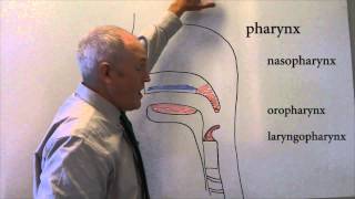
posterior pharyngeal wall positive pressure applied to the bolas tail and into these cervical esophagus where the bolus progresses to the stomach let's look at this on floor oh because that's what you're going to see in the clinic so again let's point out structure so here we are lips this is entry of the bolus from a cup these are the teeth this is the oral tongue this is the tongue base that goes all the way to the pit of the vallecula this is the hard palate soft palate nasal pharynx would be here right oral pharynx would
be here hypopharynx here this is your hyoid this is your epiglottis these are the area epiglottis here this white space here is the glottis the space between the vocal folds your written oins at this mound right here remember you have two of them you have two thinking this is two-dimensional but you have to think three-dimensional this is the posterior pharyngeal wall your PE segment is going to be here cervical vertebra five six you have your airway here your trachea and your esophagus is going to be here so let's let's play it through so here the
bolus is in the oral cavity you see that this patient has great ability to shape the bolus elevate the tongue seal the bolus to the palate anteriorly laterally posterior lis you don't see any of the contrast falling into the floor of mouth into the airway out the lips for example the fleshy part of the lips are here as the head of the bolus starts to move back your palate starts to move up and what I want you to keep a good watch on here is the hyoid bone okay when you first put something in your
mouth you will often see a little movement of the hyoid bone that's because of contraction of the suprahyoid muscles to create a stable floor of mouth for efficient tongue movement don't confuse that with onset of the fringe will swallow it's tricky it takes practice okay you'll also have movement of the hyoid when you chew but at the onset of the fringes swallow here's the head of the bolus this is the first hyoid movement on this patient right here the head of the bolus is again not only in the vallecula but it's over the epiglottis you
see that so it's beyond the vallecula and you will learn that - saliva this is actually a score of two on the NBS iymp your epiglottis is still horizontal here in fact the tip is still up and at this point your epiglottis is horizontal it's hard because this is a larger volume bolus the epiglottis tip is still up the head of the bolus is now we're in the middle of the fringe I'll swallow the head of the bolus is in the piriform sinus but see this really nice closure of the laryngeal vestibule because what's happened
is the original cartilage here have moved forward and their budding against the base of the epiglottis as the epiglottis itself goes to a horizontal position because subtraction of the the hyoid here and so you get really nice closure and you're getting first opening of the PE segment the head of the bolus is here but you can see the tail of the bolus is still in the oral pharynx which really speaks to the fact that there's this synergy of swallowing it's not just a discrete kind of phase phenomena there's much overlap there's much synergy and at
this point you have nice complete opening of the PE segment it's fairly symmetrical anterior posterior you don't see any figure-eight kind of configuration and now you're going to start to see this engagement or forward movement of the superior constrictor muscles you can see them here this is difficult to look at if you haven't been looking at it and then that movement progresses and pushes against the volus tail all the way through the PE segment
Related Videos
23:00
Swallowing
Dr. John Campbell
179,947 views

21:42
Swallowing anatomy (pharynx)
Sam Webster
147,150 views
2:46
Swallowing postures and maneuvers with vid...
Ianessa Humbert
69,559 views
13:16
How to Remember Every Muscle in the Head a...
Corporis
453,822 views
7:00
What is Dysphagia (Difficulty Swallowing)?
FreeMedEducation
359,915 views

8:05
Scared Straight – SNL50
Saturday Night Live
1,940,101 views

44:04
Gastrointestinal | Salivation: Parotid, Su...
Ninja Nerd
498,288 views

25:06
Why Standardize the MBS Study? The MBSImP ...
NorthernSpeech
80,606 views

16:37
Swallowing Mechanism: Three phases
Dr Matt & Dr Mike
85,495 views

18:25
Stroke Education - Dysphagia
Mackenzie Health
87,570 views

7:29
Dyslexia Test
Arije-Aike de Haas
2,667,016 views

18:18
Cranial Nerve BASICS - The 12 cranial nerv...
ICU Advantage
1,585,112 views

9:46
Audience Q&A – SNL50
Saturday Night Live
1,142,052 views

22:49
Larynx anatomy
Sam Webster
570,903 views

9:21
Swallowing Trouble 101
Laryngopedia
323,777 views

5:04
Exercises and Postures to Help with Swallo...
Fauquier ENT
15,321 views

48:21
Gastrointestinal | Deglutition (Swallowing)
Ninja Nerd
379,171 views

30:39
Larynx - Membranes, ligaments and muscles ...
Kenhub - Learn Human Anatomy
480,758 views
5:38
Swallowing Exercises and Postures (dysphag...
Sierra Speech
380,025 views

26:59
Dysphagia e-learning
Somerset NHS Foundation Trust
10,083 views