1) Cell Culture Tutorial - An Introduction
343.65k views1121 WordsCopy TextShare

Applied Biological Materials - abm
What is Cell Culture?
➜ Cell culture is an incredibly useful in vitro tool in cell biology research...
Video Transcript:
In biology, cell culture is the process by which cells are grown in controlled conditions outside of its native environment. This technique has enabled scientists to use cells, the fundamental unit of life, as an imperative model system for understanding physiological processes in the human body. One of the major contributions of cell culturing is the development of polio vaccines; back in the early 1930s, researchers had to use live animals to grow poliovirus, but with cell culture, researchers were able to have greater control of virus production which eventually led to the creation of the vaccine.
Fast forward to the 1970s, mammalian cell culture was used to create properly folded and glycosilated proteins, such as interferons and antibodies, that were impossible to make in prokaryotic E. coli cells because E. coli cells lack post-translational modifications.
Nowadays cell culture is being used widely in laboratory settings, particularly in the field of vaccine research, cancer research, and protein therapeutics. Cells that are used in laboratories can be generalized into two categories: primary cells and established cell lines. Primary cells are cells that are directly prepared from an organism’s tissues.
If grown under the right conditions, primary cells will grow and proliferate, but they are only able to do so a finite number of times. This is because each time as the cells divide, part of its DNA’s telomere is lost and after a certain number of cell divisions, the cells would reach senescence when the cells can no longer divide. Established cell lines, on the other hand, are able to escape the normal constraints of the cell cycle and grow indefinitely, making them extremely useful for long-term research.
Whereas primary cells are obtained directly from donor tissues, cell lines can be derived from clinical tumors, or created from transforming primary cells with viral oncogenes or chemical treatments. These reagents or treatments target one of the checkpoints in the five phases in the mammalian cell cycle. Checkpoints are there to ensure that the cell meets the appropriate requirements in order to move on to the next stage of making daughter cells.
One of the checkpoints is at the end of G1, which allows cells to commit to cell division. The second checkpoint is at the S-phase, and is there to check the quality of the replicated DNA and to determine if any DNA repair mechanisms need to be activated. The third checkpoint is after G2, and its purpose is, again, to ensure that DNA has been replicated completely and is undamaged.
Finally, there are several checkpoints within mitosis to ensure that the cell is in a proper position to complete cell division. When a cell starts to divide uncontrollably, it is because they have evaded these checkpoints and the cell cycle is allowed to proceed and cells continue to proliferate. Many viral oncogenes, such as E6 and E7 from the human papillomavirus, can degrade controllers of the cell cycle; namely the tumor suppressor genes, to turn a normal cell cancerous.
Primary cells and established cell lines in culture usually adopt one of the two growing forms: they either grow as monolayers or are free-floating in culture media. Cells that form monolayers are also often referred to as adherent cultures while the free-floating cells are called suspension cultures. Knowing the growth properties of the cells can help scientists choose the appropriate cultureware for their experiments.
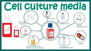
To keep the cells alive, it is important to keep the conditions as close to the physiological conditions as possible, therefore supplements such as amino acids, inorganic salts, and vitamins, are added into the culture media. Traditionally other vital supplements including macromolecules, lipids, and growth factors are supplemented in the form of fetal bovine serum (FBS). However, there are a lot of uncertainties with regards to the use of FBS.
For example, as scientists do not know the serum’s exact composition, it is always subjected to lot-to-lot variation and this in turn, can affect the cultures. Recently, defined media, where the exact composition of the media is known, are on the rise to address this issue. As physiological fluids have a neutral pH, the culture media also have a buffering system to ensure that the proper pH is maintained.
One way to achieve this is to use sodium bicarbonate to keep the pH of the media between 7. 2 and 7. 4 with 5-10% gaseous carbon dioxide.
Another common buffering system involves the use of zwitterions, such as HEPES. In both kinds, it is common to have a pH indicator such as phenol red added to the media so the scientists can visually monitor the changes in pH. Different cell lines generally require different types of media, and the most commonly used commercial media today are Dulbecco’s Modified Eagle Medium (DMEM), Roswell Park Memorial Institute-1640 (RPMI), and Ham’s F12 Nutrient Mixture (F12).
Apart from using the appropriate media for growth, the cells are also maintained in the correct temperature. For example, most mammalian cells are incubated at 37 degrees Celsius for optimal growth, while cells derived from cold-blooded animals should be kept at lower temperatures. Other measures to take when working with cell cultures is to ensure that the cells are kept under a sterile environment.
Microbial contaminates such as bacteria, fungi, or mycoplasma can ruin the culture and lead to invalid experimental data. A Laminar flow hood is always used for cell culture work. It maintains a steady, clean airflow and prevents outside contaminants from entering the culture.
In addition to microbial contaminations, the cells can also be contaminated by other cells. Cell lines can be overgrown and replaced by other fast-growing cells inadvertently introduced into the original culture. The consequence of using a misidentified cell line is making false scientific conclusions.
Authenticating cell lines using Short-Tandem Repeat profiling, or STR is recommended for cultures from time to time. abm provides a wide range of cell culture related products, ranging from the starting materials, such as primary and immortalized primary cells, to immortalizing agents, media and plastic ware. We also have specialized PCR-based detection kits and drug treatments for mycoplasma contamination.
Lastly, since many scientific journals and funding agencies now require evidence of cell line authentication by STR profiling as a requisite for publishing or obtaining funding, we are proud to now offer this service to our customers. Cell culture technique has advanced our scientific knowledge since the 1930s and it continues to provide us valuable information about all aspects of biology. If you’d like to read more, please visit our knowledge base at the link below.
Also, be sure to check out our next video for cell culture techniques. Please leave your questions and comments below, and we will answer them as soon as possible. Thank you for watching.
Related Videos
10:26
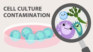
Cell Culture Contamination and Quality Con...
Applied Biological Materials - abm
17,926 views

23:37
Mammalian cell culture
Neil Cross
107,361 views

9:15
2) Cell Culture - Recombinant Adenovirus E...
Applied Biological Materials - abm
100,294 views
9:16
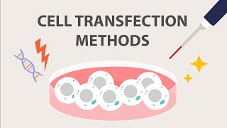
Introduction to Cell Transfection: Part 2
Applied Biological Materials - abm
2,725 views

6:13
Cell Immortalization: How to Immortalize C...
Applied Biological Materials - abm
54,652 views
5:23
Passaging Cells: Cell Culture Basics
Thermo Fisher Scientific
944,072 views
16:49
Introduction to Cell Culture
The Ahmed Lab: Northwestern Neurosurgery
11,243 views
7:00
The Basics of the Recombinant Lentivirus S...
Applied Biological Materials - abm
191,083 views
9:27
Animal cell culture media
Animated biology With arpan
92,268 views
9:10
How to Screen for CRISPR Knock Out Cells
Applied Biological Materials - abm
577 views
4:34
Cell Culture: Cell Culture Basics
Thermo Fisher Scientific
430,220 views
6:39
Introduction to Cell Transfection: Part 1
Applied Biological Materials - abm
5,552 views
5:08
Aseptic Techniques: Cell Culture Basics
Thermo Fisher Scientific
600,028 views
5:50
Tissue Culture Series #1: How to Thaw Cell...
MilliporeSigma
14,909 views
9:36
Lentivirus Transduction Protocol: Infectin...
Applied Biological Materials - abm
44,122 views
8:53
Cell Culture (Attached Cell)
Abnova
242,271 views
9:21
Adenovirus Production: How to Clone, Packa...
Applied Biological Materials - abm
9,281 views
13:29
Introduction to cell culture, splitting ce...
Dr Germán Rosas-Acosta
110,157 views

6:49
Sub-culturing cells || Cell culture basics
Animated biology With arpan
45,837 views
6:08
The Basics of Lentivirus Production/Packag...
Applied Biological Materials - abm
68,713 views