Cardiac Conduction System and Understanding ECG, Animation.
6.99M views410 WordsCopy TextShare

Alila Medical Media
The cardiac conduction system explained clearly and simply.
Please NOTE: this video talks about PQ...
Video Transcript:
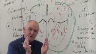
The cardiac conduction system consists of the following components: - The sinoatrial node, or SA node, located in the right atrium near the entrance of the superior vena cava. This is the natural pacemaker of the heart. It initiates all heartbeat and determines heart rate.
Electrical impulses from the SA node spread throughout both atria and stimulate them to contract. - The atrioventricular node, or AV node, located on the other side of the right atrium, near the AV valve. The AV node serves as electrical gateway to the ventricles.
It delays the passage of electrical impulses to the ventricles. This delay is to ensure that the atria have ejected all the blood into the ventricles before the ventricles contract. - The AV node receives signals from the SA node and passes them onto the atrioventricular bundle - AV bundle or bundle of His.
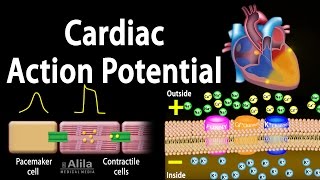
- This bundle is divided into right and left bundle branches which conduct the impulses toward the apex of the heart. The signals are then passed onto Purkinje fibers, turning upward and spreading throughout the ventricular myocardium. Electrical activities of the heart can be recorded in the form of electrocardiogram, ECG or EKG.
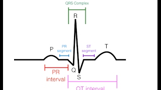
An ECG is a composite recording of all the action potentials produced by the nodes and the cells of the myocardium. Each wave or segment of the ECG corresponds to a certain event of the cardiac electrical cycle. When the atria are full of blood, the SA node fires, electrical signals spread throughout the atria and cause them to depolarize.
This is represented by the P wave on the ECG. Atrial contraction, or atrial systole starts about 100 milliseconds after the P wave begins. The P-Q segment represents the time the signals travel from the SA node to the AV node.
The QRS complex marks the firing of the AV node and represents ventricular depolarization: - Q wave corresponds to depolarization of the interventricular septum. - R wave is produced by depolarization of the main mass of the ventricles. - S wave represents the last phase of ventricular depolarization at the base of the heart.
- Atrial repolarization also occurs during this time but the signal is obscured by the large QRS complex. The S-T segment reflects the plateau in the myocardial action potential. This is when the ventricles contract and pump blood.
The T wave represents ventricular repolarization immediately before ventricular relaxation, or ventricular diastole. The cycle repeats itself with every heartbeat.
Related Videos

4:06
Cardiac Conduction System and Understandin...
Alila Medical Media
63,397 views

17:46
Heart Conduction System & ECG (EKG)
Siebert Science
127,479 views
12:24
EKG/ECG Interpretation (Basic) : Easy and ...
MINT Nursing
6,263,693 views
7:50
Cardiac Action Potential, Animation.
Alila Medical Media
2,594,546 views

18:31
Understanding Arrhythmias
Zero To Finals
120,307 views

20:44
How to Read an ECG | ECG Interpretation | ...
Geeky Medics
341,886 views

29:44
From Basics of 12 Lead ECG to How Waves ar...
Nonstop Neuron
271,523 views

15:29
Coronary circulation of the heart
The Noted Anatomist
1,646,110 views

1:19:14
EKG Basics | How to Read & Interpret EKGs:...
Ninja Nerd
2,713,384 views

23:27
Cardiovascular System 3, Heart, electrical...
Dr. John Campbell
761,859 views

3:11:52
Diagnosis of SVT in the EP lab
Dr. Joshua Cooper - Arrhythmia Education
117,844 views

48:01
Cardiovascular | Electrophysiology | Intri...
Ninja Nerd
1,591,862 views

4:23
The Difference Between Cardiac Arrest, Hea...
Health Decide
1,489,304 views

14:26
How the Heart Works (Animation)
Thomas Schwenke
134,296 views
21:33
Cardiovascular System 1, Heart, Structure ...
Dr. John Campbell
6,282,327 views

26:57
ECG Interpretation Made Easy; How to read ...
MedNerd - Dr. Waqas Fazal
683,080 views

8:43
The Cardiac Cycle is SO EASY! Stop Making ...
Interactive Biology
1,205,721 views

9:28
Heart Conduction & ECG (EKG) Interpretation
Dr Matt & Dr Mike
245,056 views

28:32
The Cardiovascular System: An Overview
Strong Medicine
732,240 views
12:14
Most Common ECG Patterns You Should Know
Rhesus Medicine
1,667,773 views