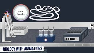
Western Blot Method - Animated Video
122.97k views1505 WordsCopy TextShare

Biology with Animations
I make animations in biology with PowerPoint, this animation video is about western blot method. Whi...
Video Transcript:
Western blotting is an important technique used in cell and molecular biology for protein separation and detection it enables the separation and identification of a specific protein of interest from a complex mixture of proteins for example a cell lysate A typical Western blot technique includes the following steps separation by size, transfer of protein to a solid support and detecting target protein using antibodies to separate the macromolecules in a sample SDS-PAGE technique can be used The first step in the SDS-Page procedure is sample preparation So that, a loading buffer containing SDS, betamercaptoethanol bromophenol blue and glycerol is
added to the protein samples the loading buffer is used in order to give all proteins present a uniform negative charge since proteins can be positively, negatively, or neutrally charged Once the loading buffer is added the samples are then heated to 95 degrees Celsius Proteins are large biomolecules, consisting of one or more long chains of amino acid residues they are formed by linking amino acids with peptide bonds and fold into specific spatial conformations driven by a number of interactions such as Hydrogen bonds, Hydrophobic interaction, Disulfide bonds, Ionic bonds. SDS is an anionic surfactant that contain a
polar head group with a net negative charge at the end of a long hydrophobic carbon chain SDS denatures the native proteins by disturbing the non-covalent forces include hydrogen-bonding, hydrophobic and ionic interactions While the reducing agent betamercaptoethanol is used to cleave the disulfide bonds Also, SDS binds fairly uniformly to the protein and the intrinsic charges of this protein become negligible when compared to the negative charges contributed by SDS this treatment brings the folded proteins down to linear molecules, with net negative charge therefore, these proteins can be separated in a polyacrylamide gel based on their molecular weight
The gel is produced by polymerisation between two glass plates anchored vertically in a cassette The gel cassette is placed vertically between two electrodes positive electrode located at the bottom of the gel whereas negative electrode is positioned at the top of the gel Next, the gel is inserted into a chamber then a running buffer is poured to allow the conduction of current through the gel SDS is also present in the gel and in the running buffer, to make sure that once the proteins are denatured, they stay that way throughout the run During separation, a molecular-weight size
marker is usually loaded onto the gel This consists of proteins of known sizes and thereby allows the estimation of the sizes of the proteins in the actual samples which migrate in parallel in different tracks of the gel Each sample is added into its own well in the gel as we have seen previously, bromophenol blue and glycerol are present in the samples bromophenol blue is a dye, that is useful for visualizing the sample in the well while, Glycerol increases the density of a sample and it is used to fall the sample into the well after the
sample application procedure, an electric field is applied across the gel causing the negatively charged proteins to migrate across the gel away from the negative electrode, and towards the positive electrode Also, Small proteins migrate relatively easily through the mesh of the gel while larger proteins are more likely to be retained and thereby migrate more slowly Due to the relatively small molecule size of bromophenol blue, it migrates faster than proteins and by optical control of the migrating-colored band the electrophoresis can be stopped before the samples have completely migrated through the gel and leave it Once the proteins
are separated, the gel cassette is removed from the electrophoresis tank Next, the glass plates are removed and, the top portion containing the wells is cut off from the gel In the next step, the separated proteins will be transferred from inside the gel to an appropriate membrane When performing a wet transfer, the gel is first equilibrated in transfer buffer so that, it is placed in a tray with the blotting buffer, on a rocking platform To make a blotting sandwich, a gel holder cassette is immersed in the transfer buffer with the black side down and the white
side up, and out of the buffer Next, a fiber foam pad is immersed in the transfer buffer Then, it is laid on the black side of the gel holder cassette After that, a piece of a filter paper is soaked in the transfer buffer Then, it is placed on top of the fiber pad once the gel has been equilibrated in the transfer buffer it is carefully placed on the blotting paper For the transfer, a nitrocellulose membrane or a polyvinylidene difluoride membrane can be used the membrane is soaked in the transfer buffer then, it is placed squarely
on the gel next, a second sheet of blotting paper is wetted with the transfer buffer Then, it is placed on top of the membrane Finally, a second fiber pad is immersed in the transfer buffer Then, it is laid on top of the blotting paper Next, the gel holder cassette is closed and locked with a clamp The most commonly used method for transferring the proteins is called electroblotting During this transfer, an electrode assembly with positive electrode and negative electrode is used The gel holder cassette is inserted into the module ensuring that the cassette is properly positioned
from negative to positive The inner module is placed into the electrophoresis chamber then, the chamber is filled with the blotting buffer Next, a lid is placed on the electrophoresis tank and electrical leads are connected to the power supply to run the blot Electroblotting uses an electric current to pull the negatively charged proteins from the gel towards the positively charged electrode, and into the membrane The proteins move from within the gel onto the membrane while maintaining the organization they had within the gel when the run is complete, the electrophoresis chamber is disassembled and the inner module
is removed Then, the gel holder cassette is removed and opened Next, starting with the fiber pad, each layer is removed until reaching the membrane After the electrotransfer of the proteins on the membrane the next step is the detection of our protein of interest using antibodies first of all, the membrane is soaked in a blocking solution which contains bovine serum albumin this step must be taken to prevent the interactions between the membrane and the antibody used for detection of the target protein once the blocking solution has been added, the membrane is incubated on a rocking platform.
The BSA protein attaches to the membrane in all places where the target proteins have not attached Thus, when the antibody is added, it cannot bind to the membrane and therefore the only available binding site is the specific target protein After the incubation is complete, the blocking solution is removed then a solution containing the primary antibody is poured into the tray next the membrane is incubated under gentle agitation The primary antibody binds to the specific target protein Following incubation, the primary antibody solution is removed then a wash buffer is poured next the membrane is incubated on
the rocking platform the membrane is washed several times in wash buffer to remove unbound primary antibody and thereby minimize background after washing the membrane, the wash buffer is removed then, a solution containing the secondary antibody is added into the tank next the membrane is incubated under gentle agitation The secondary antibody recognises and binds to a specific portion of the primary antibody Following incubation, the secondary antibody solution is removed then, the wash buffer is poured into the tank next the membrane is incubated on the rocking platform the membrane is washed several times in wash buffer to
remove unbound secondary antibodies It is crucial to thoroughly wash the membrane at this step After rinsing the membrane to remove unbound secondary antibody the membrane is incubated in a solution containing the substrate. Chemiluminescent western blot detection is the most frequently used method to detect proteins on Western blots To allow detection of the target protein the secondary antibody is commonly linked to a reporter enzyme such as horseradish peroxidase The most popular Western blot substrates are luminol-based in the presence of a hydrogen peroxide, Horseradish peroxidase enzyme catalyses the oxidation of luminol to excited state product called 3-aminophthalate
This product decays to a lower energy state by releasing photons of light at 425 nm the production of luminescence is proportional to the amount of horseradish peroxidase-conjugated secondary antibody and therefore, indirectly measures the presence of the target protein After incubation, the membrane is placed in a clear plastic wrap to prevent drying The membrane is analysed by densitometry where the resulting light signal is detected by CCD camera which captures a digital image of the western blot according to the obtained bands, we can determine the approximate size of our protein of interest as well as its presence
or absence in each sample
Related Videos
10:01
Southern Blot Method - Animated Video
Biology with Animations
203,698 views

12:40
Western Blot Protocol
SENS Research Foundation
52,434 views

8:38
SDS-PAGE, Sodium Dodecyl Sulfate–PolyAcryl...
Biology with Animations
541,340 views

18:00
Western blotting technique | principle and...
Shomu's Biology
444,577 views

7:20
DNA animation (2002-2014) by Drew Berry an...
WEHImovies
5,707,936 views

9:11
Indirect ELISA Test - Animated Video
Biology with Animations
26,067 views

7:53
Western blot protocol video
Abcam
117,146 views
16:39
Western blot explained in details | Applic...
Animated biology With arpan
14,950 views

4:23
Western Blot / Protein Immunoblot explained
Henrik's Lab
315,736 views

8:11
Northern Blot Method - Animated Video
Biology with Animations
60,323 views

58:07
Real-Time PCR in Action
USDA Animal and Plant Health Inspection Service
312,862 views

9:38
Western Blotting Protocol
Cell Signaling Technology, Inc.
160,851 views

8:02
Competitive ELISA Test - Animated Video
Biology with Animations
17,153 views

13:16
Recombinant DNA Technology - Animated Video
Biology with Animations
39,360 views

10:56
Western Blotting (Immunoblotting) : Princi...
Frank Lectures
361,183 views

6:26
Southern Blot
Abnova
174,890 views

9:57
Western Blotting
Bio-Rad Laboratories
1,072,357 views

5:27
Agarose Gel Electrophoresis - Animated Video
Biology with Animations
127,616 views

14:37
Western Blot (WB) Visual Protocol
NovusBiologicals
242,263 views

13:44
Protein Purification
Cube Biotech (Cube Biotech)
80,609 views