How to prepare the perfect Gram stain - Gram staining procedure
260.4k views1084 WordsCopy TextShare

Hardy Diagnostics
Hardy Diagnostics is your complete Microbiology supplier. To learn more about Hardy Diagnostics vis...
Video Transcript:
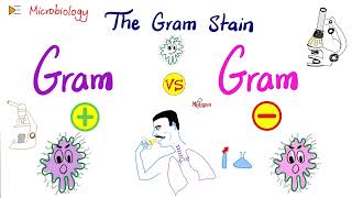
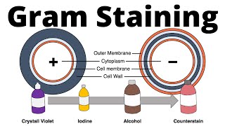
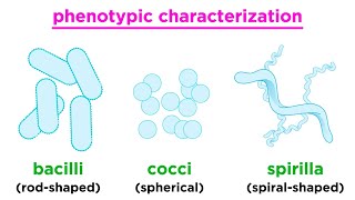
[Music] the gram stain is named after its inventor hans christian graham as a danish botanist and teacher of pharmacology he devised this important method of differentiating bacteria in 1884 that is still in common usage today the gram stain is used to distinguish between gram-positive and gram-negative cells it is the most commonly used method to observe bacteria the gram stain provides two important features the gram reaction or color of the cell and the cell morphology the shape of the cell bacterial cells can be spherical rod or corkscrew in shape and nearly all clinically significant bacteria can
be differentiated into these three groups knowing which type is causing an infection is critical to patient care the gram stain differentiates cells based upon differences in the structure of the cell wall the cell wall is made up of peptidoglycan which consists of sugars and amino acids that form a layer around the plasma membrane of most bacteria bacteria that stain dark blue purple are called gram-positive they have a thick potato glycan layer that retains the primary dye conversely bacteria that stain pink to red are called gram-negative they have a thin peptidoglycan layer that loses the primary
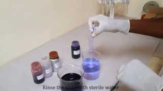
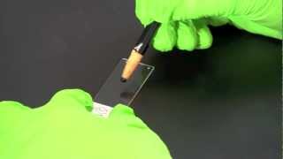
dye when flooded with alcohol the gram stain begins by adding one drop of sterile saline or deionized water onto the center of the slide then take a sterile loop and touch the edge of an isolated colony on the petri plate place the loop into the drop on the slide and swirl it around to distribute the cells evenly so the cells are one layer thick using cells from a single colony is important as this should represent only one cell type rather than a mixture of cells ideally the culture should be between 18 and 24 hours old
because older cells may show variable results alternatively you can use a collection swab from a clinical specimen or environmental sample do not heat the slide wait for the sample to air dry bacterial cells are then fixed to the slide using methanol fixing cells is important as it prevents the cells from washing off during the staining process methanol fixation is recommended rather than heat fixation as methanol preserves the cell's morphology heating the slide will cause cell distortion could increase cell debris and may cause erroneous gram reactions place or hold the slide over a paper towel and
flood the slide with absolute methanol for two minutes alternatively you may dip the slide into a copeland jar filled with methanol once two minutes have passed tilt the slide and drain off the excess methanol and let the slide air dry do not wipe or blot the slide as this can remove cells do not heat the slide cover the slide with the primary stain crystal violet for one minute crystal violet is a dark blue to purple dye that stains all cells rinse the slide with deionized or tap water to remove excess crystal violet flood the slide
with grams iodine for one minute the negatively charged iodine molecules act as a mordant and linked to the positively charged crystal violet this traps the crystal violet in the peptidoglycan layer once one minute is up rinse off the grams iodine with deionized or tap water [Music] tilt the slide at an angle over the sink or tray and add the decolorizer dropwise until the violet color stops running when the solution runs clear immediately rinse the sly with deionized or tap water to neutralize the decolorizer the decolorizer is composed of a ratio of alcohol to acetone the
ratio is normally a personal preference with faster decolorizers containing higher concentrations of acetone and slower ones containing higher concentrations of alcohol when decolorizer is added to the smear lipids are extracted from the cell wall of gram-negative bacteria lipid extraction causes an increase in cell wall permeability and the cell becomes leaky resulting in the loss of the purple dimorton complex from the thin peptidoglycan layer in contrast the effect of decolorizer on gram-positive bacteria is dehydration this decreases the cell wall permeability causing gram-positive cells to retain the crystal violet grams iodine complex note that a thick smear
will require more decolorization than a thin smear be sure to stop the decolorization process immediately after the blue dye stops running off the slide beware over decolorization can result in false gram negative results beware under decolorization can result in false gram-positive results in essence gram-positive cells will be blue purple and gram-negative cells will be colorless after decolorization the final step utilizes a counter stain of a different color to differentiate gram-negative from gram-positive cells cover the slide with saffron or carbo-fusion for one minute the gram-negative bacteria will absorb this dye causing them to stain pink to
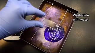
red there will be no effect to the gram-positive cells since they retain the initial crystal violet stain rinse the slide with deionized or tap water [Music] do not wash the slide excessively allow the slide to air dry by tilting it onto a paper towel or over a sink and allowing it to air dry alternatively gently dry the slide by blotting it using a lint-free bibulus paper do not use a wiping motion for this can remove the smear to summarize crystal violet exposure 30 to 60 seconds iodine exposure 30 to 60 seconds acetone alcohol decolorizer only
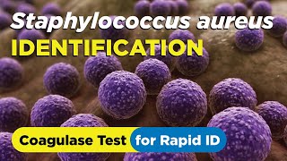
enough time to rinse away the blue stain saffron and exposure 30 to 60 seconds the slide can now be viewed under the microscope first focus on the image using the high dry objective lens marked 40x then without removing the slide switch to the high power oil immersion objective lens that is marked 100x this will result in an overall magnification of 1000x here you see two organisms routinely used for quality inspection and hardy diagnostics the purple gram positive cockeye are staph aureus the pink gram negative rods are e coli hardy diagnostics offers an improved formulation of
gram stain called gram stain advanced this kit produces superior stain results with bright vivid colors even for bacteria that are difficult to stain to test the quality of your stain and staining technique hardy diagnostics offers quality control slides the q-slide gram that are pre-inoculated and methanol fixed with gram-negative and positive bacteria thank you for watching this graham stained demonstration hearty diagnostics is your complete one-stop shop for all your microbiology supplies give us a call and experience our culture of service
Related Videos

7:51
How to Prepare a batch of Petri Plates fro...
Hardy Diagnostics
144,821 views

12:11
GRAM STAINING | Bacterial Staining Techniq...
Microbiology & Biotechnology
839,967 views

2:43
How to Perform the Catalase Test - Staphyl...
Hardy Diagnostics
67,915 views
8:09
Gram staining for differentiating bacteria...
Genomics Lab
953,622 views

11:30
Microscope Parts and Functions | How to Us...
Manocha Academy
894,422 views
17:22
The Gram Stain (Gram-Positive vs Gram-Nega...
Medicosis Perfectionalis
221,335 views
22:23
The Quest To Make Unbreakable Glass
Veritasium
2,127,838 views
10:19
How to do a hematology stain (Wright-Giems...
Hardy Diagnostics
53,921 views

6:54
Four Quadrant Streak procedure - How to pr...
Hardy Diagnostics
544,450 views
2:58
Gram positive and gram negative bacteria (...
Henrik's Lab
165,049 views

1:04:37
Bacteria | Structure and Function
Ninja Nerd
634,252 views
6:38
How to identify Staphylococcus aureus usin...
Hardy Diagnostics
17,082 views
12:56
Taxonomy of Bacteria: Identification and C...
Professor Dave Explains
394,894 views
4:28
Gram Staining
Bio-Rad Laboratories
2,007,107 views

12:57
ACID FAST STAINING | Bacterial Staining Te...
Microbiology & Biotechnology
339,676 views

9:19
Gram Positive vs. Gram Negative Bacteria
Beverly Biology
1,169,394 views

3:19:42
Rapid Revision Microbiology with Dr. Salma...
Dr.G Bhanu Prakash Animated Medical Videos
68,096 views
5:09
How to do an Acid-Fast Stain - Instruction...
Hardy Diagnostics
26,772 views
2:34
The Gram Stain Procedure
Sci- Inspi
128,481 views
14:33
Malaria Microscopy - A Step by Step Guide
MCD Global Health
250,681 views