Understanding Arrhythmias
181.64k views2059 WordsCopy TextShare

Zero To Finals
This video contains a visual explanation of various arrhythmias, aimed at helping students of medici...
Video Transcript:
[Music] [Applause] [Music] hi this is Tom from zerof finals. com in this video I'm going to be going through arhythmia and you can find written notes on this topic at zerof finals. com arithm or in the Cardiology section of the zero finals medicine book and you can find flashcards and questions to train your knowledge and help you remember the information for longer at members.
zerof finals. com so let's jump straight in aymas are abnormal heart rhythms they result from an interruption to the normal electrical signals that coordinate the contraction of the heart muscle there are several types of arhythmia each with different causes and different management options this section is a summary to help with your exam preparation and is based on the guidelines from the resuscitation Council UK from 2021 attend the relevant courses follow full guidelines and involve experienced seniors when you're treating patients let's start by talking about the cardiac arrest rhythms there are four possible rhythms that can occur in a pulseless patient these rhythms are either shockable meaning that defibr ation may be effective or nonshockable meaning that defibrillation will not be effective The shockable rhythms are ventricular tachicardia and ventricular fibrillation the two non-shockable rhythms are asy which is where there's no significant electrical activity or pulseless electrical activity which is all electrical activity except ventricular fibrillation or ventricular tachicardia and this includes if the patient has sinus rhythm but no pulse next let's talk about narrow complex tachicardia narrow complex tachicardia refers to a fast heart rate with a QRS complex duration of less than 0. 12 seconds on a normal 25 mm perss ECG 0.
12 seconds equals three small squares therefore the QRS complex will fit within three small squares in a narrow complex tachicardia there are four main differentials of a narrow complex tardia sinus tardia and the treatment of this focuses on the underlying cause supraventricular tachicardia which is treated with vagal Maneuvers and adenosine atrial fibrillation which is treated with rate control or Rhythm control and atrial flutter which is treated with rate control or Rhythm control similar to atrial fibrillation and we'll talk in more detail about atrial flutter later patients with a narrow complex tachicardia with life threatening features such as loss of consciousness or Syncopy heart muscle eseme I for example with chest pain shock or severe heart failure are treated with synchronized DC cardio version under sedation or a general anesthetic intravenous am odone is added if the initial DC shocks are unsuccessful next let's talk about broad complex tacki cardia broad complex Tachi cardia refers to a fast heart rate with a QRS comp Le Lex duration of more than 0. 12 seconds or three small squares on an ECG the resuscitation guidelines break down broad complex tachicardia into four main groups ventricular tachicardia or unknown cause which is treated with IV amone polymorphic ventricular tachicardia such as tsad deps which is treated with IV magnesium atrial fibrillation with a bundle branch block which is treated as atrial fibrillation and super ventricular tachicardia with a bundle branch block and this is treated as supr ventricular tachicardia patients with lifethreatening features should be treated with synchronized DC cardio version under sedation or a general anesthetic and intravenous amod drone is added if the initial DC shocks are unsuccessful let's talk in more detail about atrial flutter normally the electrical signal passes through the Atria once stimulating a contraction then it disappears through the atrio ventricular node into the ventricles atrial flutter is caused by a reentrant rhythm in either Atrium the electrical signal recirculates in a self perpetu in Loop due to an extra electrical pathway in the Atria the signal goes round and round in the Atria without interruption causing an atrial rate of around 300 beats per minute the signal does not usually enter the ventricles on every lap due to the long refractory period of the atrioventricular node this often results in two atrial contractions for for every one ventricular contraction which is 2 to one conduction through the atrio ventricular node and this gives a ventricular rate of 150 beats per minute there may be 3: one 4:1 or variable conduction ratios atrial flutter gives a sore to appearance on an ECG describing a similar appearance to The Cutting Edge of of a handsaw with repeated p waves occurring at around 300 per minute with a narrow complex tachicardia treatment of atrial flutter is similar to atrial fibrillation including anti-coagulation to reduce the risk of stroke based on the Chad's vascor radio frequency ablation of the re-entrant Rhythm can be a permanent solution let's talk about prolonged Q interval the QT interval is from the start of the QRS complex to the end of the t-wave the corrected QT interval or QTC estimates the QT interval if the heart rate were 60 beats per minute and this is prolonged at more than 440 milliseconds in men or 460 milliseconds in women a prolonged QT interval represents prolonged repolarization of the heart muscle cells or myocytes after a contraction depolarization is the electrical process that leads to Heart contraction repolarization is the recovery period before the muscle cells are ready to depolarize again waiting a long time for repolarization which is what happens with a prolonged QT interval can result in spontaneous depolarization in some of the muscle cells these abnormal spontaneous depolarizations that occur before repolarization are known as after depolarizations these after depolarizations spread throughout the ventricles causing a contraction before proper repolarization when this leads to recurrent contractions without normal repolarization it's called tosar de points tsad de points is a type of polymorphic ventricular tachicardia it translates from French as twisting of the spikes describing the ECG characteristics on an ECG it looks like standard ventricular tachicardia but with the appearance that the QRS complex is twisting around the Baseline the height of the QRS complexes gets progressively smaller then larger then smaller and so on to points will terminate spontaneously and revert back to sinus rhythm or it will progress to ventricular tachicardia and ventricular tachicardia can lead to Cardiac Arrest the causes of a prolonged QT interval include Long QT syndrome which is an inherited condition medications such as antipsychotics Citalopram fide sool am odone and maide antibiotics and electrolyte imbalances such as hypo calmia or a low potassium hypomagnesemia or a low magnesium and hypocalcemia or a low calcium management of a prolonged QT interval involves stopping and avoiding medications that prolong the QT interval correcting electrolyte disturbances using beta blockers but not sool and pacemakers or implantable cardioverter defibrillators acute management of tsad De points involves correcting the underlying cause for example electrolyte disturbances or medications a magnesium infusion even if they have a normal magnes nesium and defibrillation if ventricular tachicardia occurs next let's talk about ventricular ectopics ventricular ectopics are premature ventricular beats caused by random electrical discharges outside the Atria patients often present complaining of random extra or missed beats they're relatively common at all ages and in healthy patients however they're more common in patients who have pre-existing heart conditions for example es schic heart disease or heart failure ventricular ectopics appear as isolated random abnormal broad QRS complexes on an otherwise normal ECG by geminy refers to when every other beat is a ventricular ectopic the ECG shows a normal beat with a p-wave QRS complex and and t-wave followed immediately by an ectopic beat then a normal beat then an ectopic and so on management involves reassurance and no treatment in otherwise healthy people with infrequent ectopics seeking specialist advice in patients with underlying heart disease frequent or concerning symptoms for example chest pain or Syncopy or a family history of heart disease or sudden death death and beta blockers are sometimes used to help manage the symptoms next let's talk about heart block first degree heart block occurs when there's delayed conduction through the atrio ventricular node despite this delayed conduction every atrial impulse leads to a ventricular contraction meaning that on the ECG every p wve is followed by a QRS complex an ECG first degree heart block presents as a PR interval greater than 0.
Related Videos
12:14
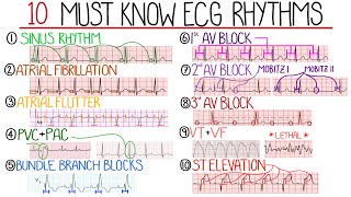
Most Common ECG Patterns You Should Know
Rhesus Medicine
1,842,658 views

1:01:14
Arrhythmias | Clinical Medicine
Ninja Nerd
245,782 views
23:43
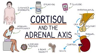
Understanding Cortisol and the Adrenal Axis
Zero To Finals
53,384 views

1:22:41
The Comprehensive ACLS Review Series!
ICU Advantage
123,500 views

17:46
Heart Conduction System & ECG (EKG)
Siebert Science
187,021 views

13:39
Understanding Thyroid Hormones
Zero To Finals
54,566 views
19:55
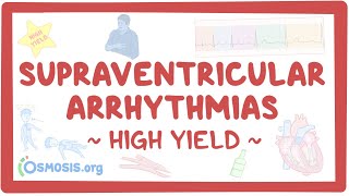
Supraventricular arrhythmias: Pathology re...
Osmosis from Elsevier
50,291 views

48:44
EKG Rhythms | ECG Heart Rhythms Explained ...
RegisteredNurseRN
1,001,139 views

19:10
Heart Murmurs and Heart Sounds: Visual Exp...
Zero To Finals
1,929,004 views

22:50
EKG/ECG Interpretation Basics Nursing NCLE...
RegisteredNurseRN
1,230,222 views
13:09
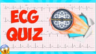
Are You A Medical Legend? Test Your Knowle...
The Learn Medicine Show
67,324 views

16:48
Understanding Cushing's Syndrome
Zero To Finals
34,971 views

18:07
Cardiac Arrest - ACLS Review
ICU Advantage
1,442,873 views
22:09
Aortic Stenosis and the TAVI Procedure: In...
Talking With Docs
50,627 views

20:44
How to Read an ECG | ECG Interpretation | ...
Geeky Medics
394,049 views

20:15
Pulmonary Function Tests (PFTs) | Clinical...
Ninja Nerd
136,152 views
16:31
Atrial Fibrillation Overview - ECG, types,...
Armando Hasudungan
379,045 views
11:04
EKG Rhythms | Top Tested NCLEX Review | Ho...
SimpleNursing
839,235 views

6:06
Cardiac Arrhythmias, Animation
Alila Medical Media
1,398,042 views

24:11
Acute Coronary Syndrome DETAILED Overview ...
Armando Hasudungan
1,377,905 views