CNS Embryology
368.13k views3867 WordsCopy TextShare

Lets Talk Medicine
This is a brief overview of the embryology and development of the central nervous system
For the sl...
Video Transcript:
[Music] hello everyone my name is Alat I'm a medical student at King bin abdulaziz University for Health Sciences and today I'm going to talk to you about the development of the central nervous system this is a brief outline to the things I'm going to talk about during the video before we start with embryology let's just start with the basics now the nervous system is divided into central nervous system and the peripheral nervous system the central nervous system is divided into the brain and the spinal cord while the peripheral nervous system is basically everything else besides
the brain and the spinal cord the brain now the brain is divided into the cerebrum which is made up of loes the temporal parietal oxop frontal lobe and also the diyon which is basically the thalamus and its family so Thalamus hypothalamus subthalamus epithalamus and then the brain stem which is made up of the midbrain the ponds and the Medela Plata and lastly the cereum this is a picture of what I just talked about so the S here the dianon here and then then you have the brain stem which is the midbrain pawns and the medel
Plata now the continuation of the mop Plata downwards here is what we call the spinal cord now let's start with some basic embriology now this is the ovary and the ovary will will release an egg now the egg will get fertilized by the sperm and then it will make up the or dividing into smaller and smaller cells and then the it will eventually give the embryo now as you can see here it's moving through the Philippian tubes reaching the uterus and it's dividing while doing that until it reaches a stage called the blastocyst so the
blastocyst is the stage that implants within the uterus now this is basically the same thing after the formation of the zot uh after fertilization you will get the embryo which will start to divide okay it will give two uh cell stage then four cell stage then it will give something called a morula as you can see here there's a membrane surrounding that embryo and this membrane it like limits the embryo from getting bigger so as you can see these cells are dividing more and more but they're smaller in size so basically the morula is just
a ball of cells which are compacted next to each other now when this P of cells start developing a cavity as you can see here we call this stage a blastocyst now the blastocyst is basically the same thing as a morula but it started developing this blast Tois cavity well but that's not the only change in the blasty now in this phase as you can see here we have different uh colors of cells you have the blue ones and the green ones now the blue cells or the cells in the middle are called the Inner
Cell mass or the embryo blast now those cells will actually give you the embryo that's why they're called the embryo blast while the cells surrounding them or the green ones are called the outer cell mass or the tropo blast and they will eventually give you the placenta now uh we won't focus on the outer cell Mouse for not interested in the placenta right now we'll only focus on the Inner Cell mass or the embryo blast so the whole embryo comes from the Inner Cell Mass so whatever differentiation is going to happen in the embryo it's
going to happen in the Inner Cell Mass the bil laminer disc now what happens here remember the blasty in the last uh picture now the Inner Cell Mass started to differentiate into two layers of cells one called the epiblast which is blue color and one called the hypoblast now those two uh epithelial layers start developing cavities within them so within the epiblast you have the amniotic cavity and within the hypoblast you have the yolac and this is what we call the bilaminar disc because it's two layered phase of the embryo one is the epiblast and
the second is the hypoblast now this is another picture of that b liner disc now I need you to imagine something this disc is not as we see it right here now basically this is just a cross-section this is a cut imagine the embryo is like this and this is where you're taking your cut so th those layers like the ippi blast and hypoblast are like here they're only one cut of the system now let's just imagine we're taking them as a disc like a real disc so this would be your epiblast and a layer
down below it would be the hypoblast now what happens to this dis just imagine with me now this dis is fill with layer cells of the AP blast right within this layer of epiblast or this plate you have two membranes okay one which is here and the other on the other side now this is the cranial end and this is the codal end okay now on the cranial end you have the Oro farial membrane which will give the future mouth of the baby and on the coral end you have the Kaka membrane which will give
the anus of the baby okay so imagine this was our plate okay now what happens in this picture as you can see here we have the epiblast the hypoblast so basically first what happens is that that some cells of the ablast will start to degenerate or or die those cells will give us what we call the Primitive streak which is just a line of empty cells that died or degenerated now and then after a while that primitive streak will even develop what we call the Primitive node so more and more cells will degenerate as we
can see here this is a primitive streak and then the Primitive node at the end of the Primitive streak which is just a bunch of cells that degenerated and then what happens after that not only did the cells die some cells from the EP Blast from here and here and here and here will start to migrate from this opening or from the streak and the note now cells migrating from the streak will give a different structure from cells migrating from the note and we'll take them one at a time in the next slide so let's
start off with the cells migrating from the Primitive streak okay this is a primitive streak and this is a primitive node now the cells migrating from the Primitive streak will start to invaginate downwards towards the hypoblast and as they go downward they start to make the layers of the embryo the other layers of the embryo so they will give you the mesoderm this is mism and then they will go towards the hypoblast and start giving you the endoderm and the cells that will stay here will transform into the ectoderm so basically what happened here all
the cells going through the Primitive streak will give you the embryon the embryonic uh like layers the endo and the Ecto and the mism and this is what we call the trilaminar disc or the three layer disc okay and now let's go to the cells coming through the Primitive node now the cells go going through the Primitive node as we can see here this is a primitive streak and this is a primitive note well actually as you can imagine they will go downwards so they're going downwards toward what toward which layer the mesoderm Imagine cells
going through the node they're going through the mism okay so first of all they're going through the mism and the second point is they don't start from the beginning as you can see this the note does not start like the streak from the beginning it starts at the end of the streak so they will start at the middle at the meod and move cranially towards the Oro farial membrane or towards the future mouth and they will make a structure as you can see here that rep it's like a line or a tube this structure is
what we call the no to cord you can also see the Noto cord here in this picture this is the Noto cord this is another picture of what we just talked about so as you can see here this is the ectoderm and uh here you can see the Noto cord forming from the node going downwards from the Primitive node and yeah the this is an not chord it began from the middle and it's also as you can see moving cranially toward the or heral membrane and also it's in in the middle layer or the mism
now what happens after that this is another picture see the not cord within the mism of the embryo you can imagine what the notocord start to do the notocord starts secreting growth factors those growth factors will act on the ectoderm above it now the ectoderm above it will start differentiating into neuro actm cells or we'll start like multiplying in the middle here and here and here and here into a uh neuroectoderm cells this is the same thing we talked about so this is the notto cord the Noto cord is secreting growth factors the uh ectoderm
above is differentiating into neuroectoderm cells getting bigger and bigger bigger and bigger until they give us something called the neural plate this is the neural plate or the neuroectoderm cells now it's basically a bunch of ectodermal cells above the above the layer the mism now what happens to this uh neural plate now as you can imagine the STC is still there so those those cells from the neural plate will start to migrate through the streak downwards towards the Miso as you can see here let's imagine our neural plate as a book okay so let's imagine
it I'm bad at drawing but bear with me this is my book it's over a table or something so this is the book we have the not cord below okay so our neurop plate as you can imagine if we have a streak here the book will start to close on itself right so as you can see when the book starts to close it will give you what we call the neural Groove cuz it's Groove downwards okay so this is a neural plate it starts to groove downwards from the streak giving us the neural uh Groove
and now some cells at the end of the book will just stay stuck in the ectoderm and this is what we call the neural folds okay and then when the groove goes downwards even more it actually joins each other the sides joins and they close giving us the neural tube and the cells that were stuck as neural folds outwards upwards will go downwards and they're they're like okay he's going to the mism I'm going too but they get cut off separately as neural Cris cells this is basically a repetition of what we just talked about
so let's just sum up okay you have the notocord notocord starts simulating the ectoderm the ectoderm forms the neural plate neural plate starts migrating toward the Primitive streak downwards when it migrates it give us gives us a Groove the edges of the groove are called the neural folds which are still in the uh ectoderm when the groove goes down it joins each other it closes as the tube and the neural folds get cut off and goes downwards towards the mism as neural Crist cells now the important thing to mention here is that this neural tube
is not empty it's filled with a CSF fold cavity so as you can imagine as this neural tube is developing into to further brain structures every part of the neural tube was will also give a cavity with it a CSF fold cavity or it will give in other words a ventricle now this is another picture of the neural tube so the neural tube has two endings a cranial end called the cranial neuropore and a codal end called the co codal neuropore now the cranial neuropore and the codal neuropore both close normally in the embryo so
the cranial neuropore closes first at day 25 and then 3 days later or at average at day 27 the codal neuropore closes failure of closure of those two neuropores will give you congenital anomalies okay so let's imagine we had our neural tube now what happens to this neural tube first this neural tube starts developing three swellings okay those swellings are called the three primary vesicles as you can see see here this tube developed one two and three swellings the first one is called the proen sephylon or the forbrain the second one is called the midbrain
or the meylon and the third one is called the romen sephylon or the hind brain so basically you need to memorize those three names your proen seon Mison and renson before you move on now what happens to those three vesicles now let's take the proin saalon now now the pro saon has two parts this part and the upper uh the lower part and the upper part now the lower part actually grows relatively slow so it will just elongate a bit as we can see in the next picture and the upper part will actually grow more
faster but it will grow a bit funny now it will go fast uh faster at the edges laterally so at the right and the left and leaving a bit of a constriction in the middle growing a bit relatively more slower so it will give you a shape like this because it's gr really fast and then a constriction in the middle and it's gr really fast in the other Edge so it will give you actually Mickey Mouse Okay a structure like Mickey Mouse now to those two structures will be called the Talon telen soplon in the
upper part so imagine telescopes are always in the upper part and then the structure that was downwards will be called the dialon so people who die are buried down in the ground so this is what I basically just talked about so let's take the first part or the proen soplon now the proen seyon as you can see here this part the lower part which is a dlon will go relatively slow and the dlon will eventually give you the thalamus and its family okay and then the upper part which is the telen soplon or the telescopes
as you can remember Mickey Mouse now Mickey Mouse will give you the cere cereal hemispheres so the petalo seal ocial whatever okay and this is for the first uh swelling or the proen sephylon now the second swelling or the meylon will actually stay the same it's in the middle it doesn't like change so it just stayed the same okay now the Robin saalon says okay Mison stays the same I'm changing into two things so it gives us two other swellings this is the first swelling and this is the first swelling and this is the second
swelling now the first swelling is called the metan sephylon and the third swelling is called the myin seyon now uh as you can see here the metan sealon will eventually give you the pawn and the cellum and the milin saalon will give you the M okay why did I put this picture here I need you to imagine how the brain looks right now to know how did it come from now as you can see this is the Talon giving you the cereum okay and then in the middle or a bit downwards to the telen sopon
you have the Dyan sealon okay giving you the thalamus and the thalamus family now then downwards you have the Meen soplon giving you the mid brain and then downwards you have the romen sealon now the romen sealon as we mentioned it has uh two parts the mlon and the milin sealon so um we mentioned the meton will give you the ponds and the cerebellum so now this makes sense because they come immediately after the mean seyon like the metan seyon the metalon comes immediately here and the second thing to mention is uh can you see
that the pawns and the cellum are actually at the same level so it just makes sense that they come from the same structure or they both come from from the Meen sephylon and then under the ponds as you can see you have the medala plung so the medala plung comes from a structure that comes under the structure that gives the ponds so the ponds came from the metal sealon the mop that will just come from the Mylon below this is a repetition of the previous slide I just need you to focus on something uh here
we're going to talk about the origin of the ventricles remember we said the neural tube is the CSF fold cavity and as it grows it will give you the cavities or the ventricles with it now just for the cavities or the ventricles imagine their position now in the normal human being like an adult human being or a born baby and just reversed the uh revers the origin back and you'll know where did they come from so uh as you can imagine the latter ventricles are actually situated within the cereal the cerebral hemispheres so they come
from the same uh origin as the cerebral hemispheres from so from the teal enlon the third ventricles are found within like between the thalamus the two thalami so they come from the same origin as the thalamus so from the D in sephylon the aqueduct of sylvus is situated within the midbrain so it comes from the structure giving the midbrain that is the Mis andalon the uh fourth ventricle however actually is uh is found within the ponds and the medulla comes from the same structure that gave the medulla and that gave the ponds which is the
mean sealon milin sealon eventually coming from the ren sealon so this is just for you to imagine so uh this is the lateral ventricle it's within the Cal hemispheres here here uh this is the third ventricle within the thalami and this is Aqueduct of cvus within the midbrain here and the fourth ventricle within the ponds and theel now the development of the spinal cord uh I need just need you to know four terminologies here so basically we first started off with the neural tube we mentioned the neural tube how does it give the the brain
now how does the neural tube give us the spinal cord basically the neural tube is aligned with epithelial cells those epithelial cells are were are what we call the neuroepithelial layer here in the middle as you can see they're also called neuroblasts now what are the neuroblasts the neuroblasts are cells capable of dividing more and more giving us um more cells outwards or more neurons so as they divide they will give you cells here on the edges okay and this layer of cells or neurons are what we call the mantle layer so the mantle layer
are new neurons or they are cell bodies in specific of those neurons so this is what we call the mantle layer as and as you can imagine if there they are cell bodies then they make up the gray matter of the SP quart and then those mantle layers will start actually giving off axons or re receiving axons depending on whether they're sensory or motor so um as you can see uh let's take the dorsal uh part now the dorsal part is actually sensory so the sensory part or the D dorsal part will actually receive axons
from outside okay and the motor port part or the part in front will actually give off motor axons now the axons going out from those cells here and here give us something called the marginal layer so the what is the marginal layer the marginal layer is axons of the cells they get myelinated and they form the white matter of the spinal cord so the mantal layer cell bodies gray matter the marginal layer the axons of those cells get melinated and they form the white matter now what is the allar plate or the AAR plate I
don't know the pronunciation and what is the basil plate it's basically parts of the mantle Zone divided according whether they are sensory or motor so anything called AAR or allar is sensory and anything called basil is just motor okay so derivatives of the neural crystals as you can imagine those other cells we said got cut off so they surround the neural tube remember we mentioned the neural tube will develop into the spinal cord and then you have this part will be sensory which is the dorsal and this is motor so the neural cryst cells are
more towards the sensory part okay and remember they are exodermal origin as we mentioned and then they will migrate laterally and give rise to the sensory gangia and this makes sense because they're more towards this part and this part will actually develop later into the spinal cord cord and then the sensory component of the spinal cord and then as you can imagine them they're near this part of the cells so they're near axons so they'll eventually give you the sympathetic neuroblast the Schwan cells pigment cells they also give aont toast and the menes and MIM
of the fial arches in this figure I just uh needed to summarize everything so you go through it uh the addition additional Point here is that you have 12 cranial nerves now where where do they actually originate from it's easy let's just memorize it you have 12 all of them uh originate from the hind brain except for four two of them will originate from the forbrain and the other two from the midbrain we mentioned the forbrain had two parts so it just makes sense that one nerve which is the first nerve Al Factory will come
from the first part of the forbrain or from the tlon and the second nerve will come from the second part of the forbrain which is the dianon and the third and fourth nerve will come from the midbrain and the rest will come from the high brain okay these are my references I'm done if you have any questions any comments just contact this email [Music]
Related Videos

26:52
Blood supply of the brain
Lets Talk Medicine
130,933 views

34:18
Embryology | Neurulation, Vesiculation, Ne...
Ninja Nerd
767,285 views

17:50
Neurulation | Neurogenesis | Neural tube f...
Animated biology With arpan
19,242 views

6:32
Gastrulation - Embryology
About Medicine
573,070 views

40 Hz Brain Activation Binaural Beats: Act...
Good Vibes - Binaural Beats
12:20
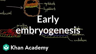
Early embryogenesis - Cleavage, blastulati...
khanacademymedicine
2,199,645 views

14:49
Embryology of the Nervous System
Dirty Medicine
78,555 views

35:16
Foundational features of the brainstem
The Noted Anatomist
367,030 views

37:36
Embryology | Gastrulation
Ninja Nerd
1,169,110 views

1:21:09
Intro to Neuroanatomy | Neurophysiology | ...
Dr. Najeeb Lectures
3,165,718 views

47:11
Head & Neck Anatomy | Embryology & Pharyng...
Mental Dental
250,139 views
12:09
Overview of the Central Nervous System (CNS)
Dr Matt & Dr Mike
505,185 views

36:42
Embryology | Development of Reproductive S...
Ninja Nerd
600,950 views

30:22
3D Embryology of Pharyngeal arches, Pharyn...
MedicoVisual - Visual Medical Lectures
144,359 views

47:41
Embryology of the Brain - Embryology
Drbeen Medical Lectures
75,398 views

35:19
Neurology | Gross Anatomy of the Spinal Co...
Ninja Nerd
1,138,173 views

12:16
The Central Nervous System: The Brain and ...
Professor Dave Explains
469,656 views

4:25
Ascending and Descending Spinal Cord Tract...
Rhesus Medicine
423,421 views
3:24
Neurulation - Animated Embryology
About Medicine
159,033 views

14:32
Embryology of the Nervous System | Develop...
Medicosis Perfectionalis
42,393 views