Western blot explained in details | Applications of western blot | CSIR NET
31.22k views2713 WordsCopy TextShare

Animated biology With arpan
For Notes, flashcards, daily quizzes, and practice questions follow
Instagram page: https://www.ins...
Video Transcript:
in this video we'll be talking about Western blot this is a detailed explanation on Western blot Western blot is a frequently used technique in a molecular biology or biochemistry lab so what does this technique detect this technique can detect specific protein or your protein of interest in a mixture of protein or a patient sample so here is a mixture of protein and our question is whether this particular pink protein or our protein of interest is present within that mixture or not Western blot is a technique which can give us this answer and the way Western
blot works is the antigen antibody interaction so here the protein of interest is detected by a primary antibody followed by a secondary antibody which is linked to some enzyme so this is why Western blot is also termed as immunoblot because it utilize the principle of antigen antibody interaction now let's talk about the Western block workflow in details so there there are broadly three sets of uh step First Step would be protein extraction from the cells or tissues or from patient serum Etc and the second step is separating the protein on a gel and the third
step is detecting the protein using antibodies in that Western blood workflow so these are the broad steps so first step is the protein extraction which is done from uh any kind of cells or tissues using lces buffer we'll talk about the lices buffer comp position in a moment second step is running the SDS Spade gel or one-dimensional gel to separate the proteins according to molecular weight and the third part is transferring the proteins all proteins from the membrane from the gel to the membrane and later on using that membrane to detect whether our protein of
interest is present or not so these are the three broad stretch steps and in each step we would focus and understand the details so the first step is basically SDS page so in this case before we even start the procedure we have to think about the Step Zero which means isolating the protein from the cells in this case proteins has to be isolated so we need to have a protein extract or protein mixture and that is done using a buffer known as ralis buffer this is the composition of the ralis buffer which has several components
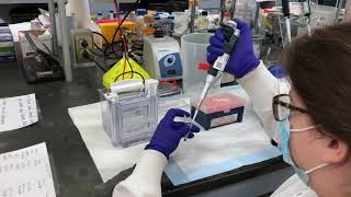
alongside this buffer a proteas inhibitor cocktail is also provided just to prevent the degradation of the proteins then next step is to run these extracted protein mixture on a SDS page one-dimensional gel so here we load the gel in the particular SDS SDS gel and then we wait for some time we allow the gel to run and the proteins are separated on basis of their molecular weight so here since we use only one parameter that is the molecular weight of the protein to separate them from each other this is known as onedimensional gel electrophoresis as
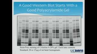
well so question is how this gel is made the gel is made out of acrylamide and NN methy bisacrylamide so these these polymerize to form the polyacryamide gel or page so in this case there is a gel holding cassette between these cassette there are two glasses where you pour the gel now the gel chemist the gel making chemistry is as follows it uses basically a free radical mechanism to polymerize Tade is one component that is used to accelerate or used as a catalyst in this process so if the free radical is represented as r dot
then the reaction goes like this described here so just remember that in uh Western blood or in case of SDS page Gil making the chemistry behind this is a free radical polymerization chemistry now the question is how the overall like Which percentage of the gel has to be made and it depends upon the need for example we want to separate a protein which is around 80 kilon or let's say 120 kilodalton and in another scenario we want to separate a protein which is like 13 kilon or 22 kilodalton so obviously we can see there is
a huge difference in the molecular weight now based on this molecular weight different types of gel or percentage of gel is used for example for bigger protein if we need to separate we used 10% gel that means it has much bigger pore size whereas in order to resolve smaller proteins we need to use a smaller por size gel like 15% gel a detailed catalog of all these percentage and which molecular ladder to be used can be available by the manufacturer company itself now let's see how proteins are actually separated in the gel we need to
talk about the chemistry and the biology behind that so Western blot sorry the SDS space gel has two components to it stacking gel and the resolving gel stacking gel is a portion where proteins are concentrated prior to their run in the resolving gel and resolving gel is the portion where the proteins are really separated on basis of their molecular weight so differences in the composition of stacking gel and resolving gel is is really important to understand and this kind of system is capable to finally resolve protein according to their molecular weight so let us talk
about the percentage of the gel so the stacking gel is broadly 5% so it has very big pore size whereas resolving gel could be variable for example in this case 12% it could be also 15 or 10% as well now the pore size is obviously larger in case of stacking gel the resolving gel has relatively smaller pore size but it also depends on the con concentration the ionic strengths are different also the pH are different so why there is so much of differences and let's try to understand that so imagine we are first uh putting
some SDS and beta markup to ethanol with the extracted protein samples so in this case what happens is if the protein has positive and negative charges when we code them with SDS it would have a uniformly negative charge around them this would allow this would ensure that when the proteins are separated on the gel they are solely separated on the base of molecular weight not the charge so now every protein is co by SDS and negatively charged now if this is the overall gel and the proteins are loaded there and they the proteins are negatively
charged so they would be moving from the negative electrode to positive electrode now there are also other components in the buffer such as the chloride ion the Glycine and also the protein which are coated with SDS so a first chlorine ion would move quickly because it's negatively charged smaller in size it would quickly move towards a positive electrode then the protein would move after that and lastly the glycine ion would move at pH 6.8 it is protonated so it is positively charged so it would be lagged behind so sandwiched between the glycine ion and the
chloride ion these protein molecules move in a stringent Manner and they all stack up before the resolving gel it's kind of kind of like a realigning the athletes near the starting line so now it's time for them to move into the resolving gel now resolving gel has a different pH always remember because it has a pH 8.8 at this point of time the chloride ion would be moving very fast now the glycin ion also start moving fast because at that particular pH it is not anymore prot alterated it has negative charge so it is now
anionic right that's why it move very quickly now the protein would be moved afterwards based on their molecular weight so this is the overall function of stacking and the resolving gel now let's the final step two is to run the gel so the gel is run around 200 Volts for about half an hour always somebody has to check for the gel while it is running and when the die front actually leaves the uh gel it is time to stop the gel after the gel is run properly it is important now to stain the gel this
is how a unstained gel broadly look like after staining with specific reagent like Kumasi blue it would look like this one can clearly understand there is a molecular weight ladder on the right hand side and there are specific bands which corresponds to different type of proteins present in this sample now the question is which stain to use and what criteria determines which stain we should be using so there are different stains which one can possibly use there is Kumasi Brilliant Blue stain there is silver stain there is ponu now silver stain is very sensitive for
example if you want to detect proteins as low as 5 to 10 nanogram silver stain is the choice Pono also detects at a level of 200 nanogram or higher now there there are other stains such as zinc stain cypro Ruby stain NY red stain now each of these stains has their own purposes own benefits and own uh disadvantages a quick overview is provided in this particular chart now let's move slowly to the Western blot because we have separated the protein in the gel now it's time to blot and detect whether our protein of interest is
present or not when we separate the gel on a when we separate the protein on a gel our protein of Interest might be present might not be present in the mixture as well it's just a separation and segregation of the protein on a gel now let's talk about how we can transfer the proteins that we have separated in the gel into a membrane and this is done by electroblot so here a pvdf membrane is placed on top of the gel and then it is sandwiched between layer of paper towels and then it is moved into
a gel holder casset further this gel holder casset is uh embedded into a buffer tank which has a transfer buffer now this transfer happens under the influence of electrical field now we already know the proteins that are present in the SDS gel they have negatively negatively charged because they are basically Co with SDS so they would possibly move towards the positive electrode right so they would move from cathode to the anode and that's what is done in this particular step So eventually all the content of the gel would be now transferred into the particular membrane
or the pvdf membrane now then a blocking step is performed it is done to prevent non-specific binding of antibodies so blocking buffer quats all the portions of the membrane that is such that the antibody does not non-specifically bind to the membrane it should find the protein of Interest always so blocking solution is generally made up of nonfat milk or Bine albine and this step ensures that antibodies later on does not stick to the membrane now then ultimately uh protein uh a primary antibody is added to detect whether our protein of interest is present or not
if the protein of interest is present primary antibody should be detecting it like shown here and if it is not present it should not be getting detected so obviously after primary antibody adding the overall solution is kept overnight in 4° centigrade in a shaking condition so later on in the next day the washing step is done to remove and wash away all the non-specific bound antibodies or other interactions later on secondary antibodies are also added secondary antibody binds on the back of primary antibod so obviously if the protein was there primary antibody should be there
and the secondary antibody should be detecting it and ultimately there would be another washing step a couple of washing steps which ensures the all the non-specific secondary antibodies bound here and there would be removed ultimately we have to develop the blot in order to develop the blot there are different strategies there are cetric strategies where the secondary antibody is bound with a enzyme which upon sub which upon giving substrate would be converted into a color product there are also chemiluminesence uh detection where there is an chemiluminiscent uh Pro converting protein which would convert a substrate
into a product which would be chemiluminiscent and then there would be also fluoresence based detection where the secondary antibody is fofor tagged So based on the f for one can detect the fluoresence coming out from that sample so all these methods can be used each methods has their own difficulties and sensitivities widely used methods are chemiluminesence and fluoresence fluoresence methods are pretty sensitive but also expensive so these gels are actually imaged in a gel scanner or blot scanner which has a Imaging system to adjust exposure Focus Etc so this is how one can get a
picture perfect Western blot now that let's now let's talk about the applications of Western blot in bit more details with some examples so here are some clinical applications and biomedical applications it's a very widely used bread and butter technique for biochemistry Labs so let's say how Western blot can be used to study cell signaling in a cell signaling pathway you always know that there are some proteins that phosphorate other Downstream proteins and this is how signaling Pathways go on so sometimes phosphorilation is a readout of activation of a signaling path pathway so in this case
how one can use Western blood to detect these kind of scenarios so one can really have antibodies again un phosphorilated versus phosphorilated conformation of that protein and try to detect a ratio of these two from the P from the particular sample so let's say there is a mapk pathway we know that there are Ras mapk pathway involves RAF make Ark Etc so phosphorilation of Ark is one of the sign that mapin pathway is active ated and this can be actually visualized using Western blot so obviously if there is more and more phosphor Arc then the
signaling pathway is activated right so in this case in the blot you can see when the liand is present there are more phospho AR compared to the normal Arc so Arc to phosphor ratio is a indicative in this particular case of the signaling activation now let's say another signaling pathway involves let's say nuclear receptors which upon Lian binding move from the cytoplas m to the nucleus so the question is how this can be detected on a western blot so obviously one can simply um look for Lian versus no liand situations and extract the nuclear and
cytoplasmic fractions using Ultra sentation from those extracts one can probe for the protein of interest in this case this particular nuclear receptor from from these extracts and if somebody sees a enhanced nuclear fraction or presence of that protein in nuclear fraction and a reduction in the cytoplasmic fraction that means what that means the protein has translocated into the nucleus in this case carefully notice the cytoplasmic fraction when the liand was absent there are more proteins in the cytoplasm than the nucleus but when the Lian was uh present the plus sign look the cytoplasmic fraction has
reduced and the nuclear fraction has increased instead that simply means the protein has physically moved from the cytoplasm to the nucleus so these kind of research questions can be addressed using Western blot these are very simple examples that I I'm providing you for your understanding so I hope this video was useful if you like this video give it a quick thumbs up don't forget to like share and subscribe you can support our Channel using super thanks which is a heart shape icon present on the bottom right corner of the video by clicking on it you
can pay via Paypal PTM or UPI see you in next [Music] video
Related Videos

6:04
RNA extraction using trizol method
Animated biology With arpan
49,647 views

12:40
Western Blot Protocol
SENS Research Foundation
74,134 views

14:27
CRISPR-Cas9 Genome Editing Technology
Professor Dave Explains
796,836 views

14:37
Western Blot (WB) Visual Protocol
NovusBiologicals
249,399 views
17:26
Signal Transduction Pathways (G-Protein, R...
Dirty Medicine
603,551 views
16:31
Introduction to the immune system
Osmosis from Elsevier
296,840 views

35:56
Western Blot Protocol Overview
The Ahmed Lab: Northwestern Neurosurgery
16,405 views

39:16
Cell Membrane Structure & Function
Ninja Nerd
452,288 views
17:41
Single cell RNA sequencing overview | ScRN...
Animated biology With arpan
43,747 views

35:15
Understanding CRISPR-Cas9
Andrew Douch
236,220 views
22:20
Protein Structure - Primary, Secondary, Te...
Medicosis Perfectionalis
101,946 views
45:42
Can We Trust Western Blots?
Bio-Rad Laboratories
49,032 views

14:37
Sanger DNA Sequencing, From Then to Now.
ClevaLab
104,617 views

16:53
Biologist Explains One Concept in 5 Levels...
WIRED
4,221,724 views
40:41
Bacterial Genetics
Ninja Nerd
363,273 views

17:59
Krebs Cycle | Made Easy!
Dr Matt & Dr Mike
354,224 views

7:53
Western blot protocol video
Abcam
145,897 views
31:35
SDS PAGE and Western Blot
Keppetipola Research Lab
46,465 views
14:10
Translation (mRNA to protein) | Biomolecul...
Khan Academy
1,320,092 views
14:41
Western Blot Protocol
Addgene
12,827 views