Trochlear Nerve - 4th Cranial Nerve (CN IV), Superior Oblique, Cavernous Sinus Syndrome - Neuro
9.8k views4560 WordsCopy TextShare

Medicosis Perfectionalis
CN IV...Trochlear Nerve...Cranial Nerve 4 (CN IV) Neuroanatomy, Superior Oblique, Cavernous Sinus Sy...
Video Transcript:
hello wonderful people it's meosis perfectus where medicine makes perfect sense welcome back to my neuroanatomy playlist in previous videos we talked about cranial nerves we talked about the AL Factory nerve which is special sensory apher then we talked about the second cranial nerve which is optic nerve also special sensory apher we also talked about the visual pathway and the diseases or the deficits that can affect that pathway and in the last video we talked about cranial nerve 3 or the oculomotor nerve which was really oculomotor motor to the eye muscles it also carried Paris sympathetic fibers with it to accommodate the lens and constrict the pupils as for today it's time for a purely somatic motor nerve known as the tro clear nerve and this is the fourth cranial nerve why do we call it tro clear nerve because it comes close to the troa hit the like button click the Subscribe button and let's get started please refer to my videos in the anatomy playlist or if you just want the neuroanatomy ones check out my neuroanatomy playlist Al Factory nerve special sensory apher for the sense of smell olfaction or smell is the only Sensation that does not relay in the thalamus before it reaches the cortex then we talked about the optic nerve special sensory aphant this is how we see but how do you move the eye then if I want to move the eye I got to use cran nerves three four and six remember the line of the sand in front is motor behind is sensory most of the time basil plate AER plate if I want to see oh seeing something looking at the tree is a sensation vision is a sensation that's why the visual cortex is behind the line but what if I want to move my eyeballs then you got to work with the frontal eye field since this is motor it is in front of the imaginary line in your brain it passes by the central sulcus during embryology it was called the sulcus limitans the same story applies for the spinal cord we'll talk about the spinal cord in detail as well as the spinal tracts white matter gray matter spinal nerves you name it after you finish the cranial nerves but you can also divide your spinal cord into two parts with the imaginary line here in front is mottor behind is sensory the brain and spinal cord are central nervous system anything else is peripheral nervous system how about the cranial nerves and the spinal nerves they are peripheral nervous system except one that one is the optic nerve the optic nerves are cranial nerve but it's not peripheral nervous system it is actually part of the central nervous system as we have discussed before and this fact has many clinical implications today we're talking about the trolear nerve this is part of the peripheral nervous system what is the structural unit of the nervous system the neuron is how about the functional unit that's the reflex arc here is the Soma or the cell body of the neuron and here's the axon a collection of Somas in the CNS is called a nucleus but a collection of Somas in the pns is called a gangion how about the axon a collection of axons in the CNS is a tract a collection of axons in the pns it's a nerve now I'll tell you today about the nucleus of the trolear nerve why do you say nucleus because it's a collection of cell bodies ins inside the central nervous system the nucleus of the trolear nerve is inside the midbrain okay and this is a collection of cell bodies in the CNS and then as the nerve exits the CNS and goes or passes anteriorly it leaves the CNS so now it's in the peripheral nervous system a collection of axons in the peripheral nervous system is called what a nerve and this will be called your trar nerve which started as a trolear nucleus recall that myin appears white in color so melinated fibers the white matter but UNM ated fibers represent the gray matter today's nerve the TR clear is motor not sensory that's why it started in the brain and then it goes outside from the brain to somewhere else I. E eent this nerve is somatic eant or general somatic eant cranial nerves one and two emerge out of the forbrain three and four emerge out of the midbrain 5 6 7 and 8 from the ponds 9 10 11 12 from the medulla and 11 borrowed another piece from the spinal cord today we're talking about the trolear nerve or the fourth cranial nerve so it emerges out of the midbrain three and four emerge from the midbrain three is from the upper part of the midbrain but four is from the lower part of the midbrain ocul motor emerges at the level of the superior calculus of the midbrain but the trolear nerve emerges at the level of the inferior culus of the midbrain brain remember that the midbrain is called what Meson in the next video you will learn about the mesencephalic nucleus guess where is the mesencephalic nucleus located it is located in the mesen cilon OR midbrain if you actually understand what these words mean you will get many questions correctly on your exam just by understanding the lingo you can download these colorful handwritten notes on my website medicosis Perfection is. com I help you under understand and pass exams the reflexes of the eye are in the midbrain and no wonder look at the oculomotor nerve and the trolear nerve they come out of the midbrain is my trolear nerve somatic or autonomic or both answer it is only somatic is it somatic motor or somatic sensory only somatic motor that's why we call it General somatic ephant or simply somatic eant does the trolear nerve contain any visceral or autonomic fibers no so no sympathetic no parasympathetic none which means there are no pre galonic or post galonic fibers because there are no ganglia and if there is no gangion for the trolear nerve you will not hear of a white Rus communicans or a gray Ramos communicans let's review the cranial nerves that we discussed before Al Factory nerve is the first cranial nerve it starts from between the mosa in the rof of my nose starting with these lovely bipolar neurons known as olfactory fibers and then we contact the mitro cells in the olfactory bulb we synapse together the olfactory bulb will take us to the olfactory tract which will take us to the telen sephylon which is part of the forbrain so it is nerve and then bulb then tract then forbrain optic nerve is very similar here is a nerve here is a bulb known as the optic kaym rather than optic bulb and then optic tract and then instead of tanyon say dialon another part of the forbrain the olfactory nerve started from the mucosa of my nose but the optic nerve starts from the gangion cell layer of my retina then the ocular motor nerve it is motor eent do you mean somatic motor or visceral motor both recall that cranial nerves 1973 i e cranial nerve 3 cranial nerve 7 cranial nerve 9 and cranial nerve 10 have parasympathetic function oculomotor is number three it has parasympathetic function cranial nerve 3 and four start in the midbrain I start here I have two nuclei one is for somatic motor known as the ocul motor nucleus or the motor nucleus of the oculomotor nerve and the second nucleus is called the Edinger Vistal nucleus which is parasympathetic and then I have nerve fibers for the oculomotor nerve which are close to the ancus of the temporal lobe as well as to the edge of the tentorium that's why unle herniation or trans tentorial herniation can ruin my oculomotor nerve then it delves into the lateral wall of the cavernous sinus some Duram action then the oculomotor fibers enter through the superior orbital fissure now we are in the orbit we will also enter through the tendonous ring will divide into Superior division of oculomotor and inferior division of oculomotor superior division will supply elevator pbbr superioris including the superior tsal muscle by borrowing some sympathetic fibers from the plexus that surrounded the internal cored artery and the Opthalmic artery I will also Supply the superior rectus muscle as for the inferior division of the oculomotor nerve I will give the inferior rectus inferior oblique and the medior rectus the nerve of inferior oblique has another function remember the edur Vistal nucleus it will take some of those fi fibers that's parasympathetic relay into the ciliary gangion then short ciliary nerves to the ciliary muscle and the constrictor pupil to accommodate my lens and constrict my pupil for near Vision who else comes out of the midbrain the trolear nerve so now we have cranial nerves three and four emerging from the midbrain the difference is the level since three comes before four you'll find that ocul motor nucle nucleus is Superior in an atomical position to the trolear nucleus what do I mean I mean that the nucleus for oculomotor nerve is at the level of the superior calculi of the midbrain however the nucleus for the trar nerve is at the level of the inferior culi of the midbrain how do cran nerve 3 ocul motor emerge one from here one from there here's right oculomotor left oculomotor and then it passes anteriorly until it leaves the midbrain conversely I want you to look at the trolear nerve here is the trolear nucleus on the right and trolear nucleus on the left look at the trolear nerve oh we're going to the back and then turning around until I leave the posterior aspect and go to the anterior aspect like this and I will go in a similar route to the oculomotor nerve which makes the trolear nerve the only one of the 12 cranial nerves that emerg from the posterior aspect of the brain instead of emerging from the anterior aspect how do I remember this here is a very improper nimonic trolear nerve with a t reminds me of the tush with a t it emerged from behind if you're more polite you can say from the tail end see that midbrain it literally looks like a pair of shorts the front of the short is called the cross cerebra and this is part of the cerebral p dcle but the back end is called what it's called the tectum which reminds me of rectum so now the trolear nerve is the only nerve that leaves from behind tush or tail end which part which behind are you talking about the tectum now let me tell you something about the pedones draw this with me please we will try to draw this so here is the cerebral cortex beautiful temporal lobe with the ancus nice nice nice and then like this okay then what I want you to add the midbrain here oky doie which looks like a pair of shorts as you have seen then what after this I have the pawns amazing followed by the midulla upang midbrain plus pawns plus medulla they look like a stem hence brain stem this is the stem of the tree and here are the leaves of the tree behind the brain stem there is the lovely cerebellum cool now let's talk about the big cables known know as pedones how does the midbrain communicate with the cerebrum by something called cerebral pedones one on the right one on the left how does the same midbrain communicate with the cerebellum behind it with Superior cerebellar ponal one on the right one on the left how does the ponds communicate with the cerebellum behind it midal cerebella ped unles one on the right one on the left how does the Mulla ablong communicate with the cerebellum by the inferior cerebella pedones one on the right and one on the left so these are cereal pedones but these are superior middle and inferior cere pedones cerebral cere pedones let's look at the midbrain from behind this is the tectum which is the hiney of the midbrain Superior culi here INF fear culi there cuz remember that the rectum I mean the tectum of the mid brain has four balls so the front of the midbrain looks like a pair of shorts the legs of the shorts the front has the two legs the back has the four balls Superior balls inferior balls the oculomotor nerve nucleus is at the level of the superior culi but the trolear nerve nucleus is at the level of the inferior culi then after emerging from those nuclei in the midbrain the lovely trolear nerves will decate some PR cross action the right will go to the left and the left will go to the right some fibers decate some fibers do not we don't care right now just remember that after it exits through the midbrain from the highe it will pierce the superior midy veum if it's called Superior midar veum you can bet that there will be an inferior midar veum downstairs and this is what this is the fourth ventricle containing cro spinal fluid now the tro clear nerve will take a turn around the superior cere peduncle which connects what to it it connects the midbrain in front with the cerebellum behind here is the left trolear nerve and here is the right trolear nerve that's it as far of the trolear nerve for now let's just review this you see the superior cicus what's the function of superior cicus o vision Bingo the cicus of vision is connected to the geniculate body of vision known as the lateral geniculate body remember my neonic lateral genicular body with an L will make me see light with an L however in falus is for hearing and it's connected to the genicular body of hearing known as the MGB pneumonic the MGB with an M will make me hear music with an M then we have a connection between Superior cicus and LGB this is called a superior brachium brachium literally means arm that's why we have the brachel archery in your arm taada and the biceps brachi and the bracho radialis and the brachio calic got it so brachia means arm here is an arm that connects the superior calculus to the LGB you can call it Superior brachium or brachium of superior calculus similarly here is an arm that connects the inferior cicus to the MGB and this is called inferior brachium or brachium of the inferior cicus why is this lateral genicular body and this medial genicular body because look literally the lateral is more lateral to the medial oh this is more lateral and this one is more medial it makes sense once you understand what the flip you're talking about we have said it before and we'll say it again if you are purely motor I mean purely somatic eent you will leave the brain stem straight forward like this and your nucleus will be more Central more towards the midline such as the ocul motor nerve especially its motor nucleus which is known as the ocul motor nucleus and the trolear nerve the trolear nerve nuclei are very close to the midline yes I know that the trolear nerve is weird it goes behind but it then comes in front and when it comes in front it is very close to the midline conversely if you are not pure blood if you're not purely motor like this if you're mixed like motor and some autonomic and maybe some brain kill maybe some sensory with it Etc then you will not be closer to the midline you will be closer to the lateral side and you tend to be more posterior compared to the fibers that leave anteriorly let's review the extraocular muscles most of them were supplied by cranial nerve 3 which is the oculomotor nerve and these included the levator PP superioris the superior rectus the inferior rectus the medior rectus and the inferior oblique muscles however there are two muscles that are not supplied by the oculomotor nerve one is the lateral rectus which abducts the eyeball if it abducts it got to be supplied by the abducent nerve and the superior oblique muscle which is supplied by the trolear nerve which is today's topic why do we call it trolear nerve because the superior oblique muscle passes by the trolea which acts as a pulley hence trolear nerve remember that the troa is medial in your orbit not lateral it is medial now to my favorite part of the electon please bring pen and paper and let's draw this together we're talking about trolear nerve so it leaves the midbrain so here is the midbrain which looks like a pair of shorts and here will be the nucleus of the trolear nerve the trolear nerve is crazy in that it leaves the midbrain from behind not from front and then wet then we are also related to the edge of the tentorium that's why trans tentorial herniation can affect the choar nerve although it affects the oculomotor more often the same ch nulear nerve will continue forwards until it reaches the lateral wall of the cavernous sinus and it will move inside the lateral wall of the cavernous sinus then it will leave the cavernous sinus and will enter through the superior orbital fisure now the trolear nerve is inside the orbit will it enter through the common tendinous ring of the reti no it will not enter the tendonous ring which is different from the oculomotor because the oculomotor did enter the tendonous ring so let's draw the tenderness ring here the choar nerve will not enter through this instead the choar nerve will go up near the trolea to supply the famous Superior oblique muscle the superior oblique muscle then attached to the eyeball here is my eyeball amazing and what does this super oblique muscle do when it contracts it depresses my eyeball it makes it look downstairs like this not just downstairs it also slightly abduct the eyeball making it look laterally as well as externally rotate the eyeball external rotation is known as extortion this movement was named by a lawyer internal rotation of the eyeball is in torsion external rotation is extortion here's the trolear nerve it is somatic motor it is eant we call it somatic ephant or general somatic eent okay I start here from the trar nucleus which is a motor nucleus in the midbrain it's by the way related to the medial longitudinal facul which makes your eyeball look medially it's called medial lenal because it makes you look medially and this is ruined in a disease known as multiple sclerosis from the chocar motor nucleus emerges the trolear nerve where is the location of the nucleus it's in the midbrain at the level of the inferior culus just anterior to the Cal Aqueduct of sylvus the trolear nerve exits from from behind takes a turn around the superior cerebellar peduncle and then it moves or passes on the lateral aspect of the midbrain just near the lateral aspect then it enters into the cavernous sinus inside its lateral wall before it enters into the cavernous sinus it was related to the edge of the tentorium of course anytime you get close to the cavernous sinus you will be related to the internal cored artery the trolear nerve enters the orbit via the superior orbital fissure then it goes to supply the superior oblique muscle it's called trolea because we are related to the trolea when the superior oblique muscle contracts it depresses the eyeball even though it's called Superior oblique yeah it depresses it makes my eye look inferiorly also abduction or lateral movement of the eyeball as well as external rotation which is known as extorsion extorsion with an S not with a T please let me know what else passed through the orbital visure did the optic nerve pass through it if the answer is no then where did the optic nerve pass through did the otor pass through it how about thear trigeminal abducent what are all the structures that pass through the superior orbital visure and what are all the structures that pass through the common tendonous ring of the reti why do we call it this name because all of your reti emerge from the common tendonous ring and I'm talking about the superior rectus inferior rectus medor rectus and later rectus to imagine the action of the superior oblique muscle just look at this here's the insertion and here's the origin just get the insertion closer to the origin as you contract this will be pulled up so the eye itself will look down that's depression this stro CA is medial but as you pull medially this way the eye itself will look laterally and this is the abduction part let's take it to the clinic we'll talk about choar nerve PSY and cavernosus thrombosis do you remember aom motor py yeah we talked about this in the previous video in oculomotor py this epyal eye will have tosis or dropy eyelid down and out as you see and I will have blown pupil or medis now let's talk about tro clear nerve Psy we'll suppose that the choar nerve that is suffering is my right tro C nerve which eye will suffer the right eye remember that the trar nerve Supply is only one muscle which is the superior oblique muscle the superior oblique muscle M she rest in peace used to depress my eye make me look downstairs oh I cannot look downwards anymore instead I will look up the superior oblique Mai rest in peace used to abduct my eye which means make me look laterally now I cannot look laterally so I will look medially like this it also used to allow me to externally rotate the eyeball like this instead I will go into internal rotation this is what you see on physical examp exam ask the patient to look here to this side follow my finger here oh this normal eye follow is no problem but this eye will not be able to follow smoothly instead it will shift upwards like this when I lose the superior oblique I look superiorly this is from the physician's perspective but how about from the patient's perspective what will be the patient's complaint if I cannot look down my biggest obstacle will be climbing downstairs or upstairs especially downstairs because notice the last time you walk downstairs if you remember you had to look down via your Superior oblique but if I do not have a functioning Superior oblique I will be unable to look down which means I will have difficulty walking downstairs many of these patients unfortunately fall while walking downstairs it's time for you to start appreciating your tro clear nerve before it's too late another clinical disease that we'll discuss briefly is cavernous sinus syndrome remember when someone punched me in the dangerous triangle of the face some of these veins will communicate with the big vein known as the cavernous sinus a sinus is a big vein such as the sinuses in your heart remember the coronary sinus the cavernous sinus the Dural Venus sinuses and what does the word cavern mean oh it's like a cave oh it's a Confluence of caves of veins like this oh that's a cavernous sinus right there and then what remember the structures that pass through through the cavernous sinus if this is my cavernous sinus remember that I had the ocula motor passing through the lateral wall I had the trolear nerve passing through the lateral wall I also have V1 and V2 the Opthalmic and the maxer branches of the trigeminal nerve passing through the lateral wall all of these symptoms will lead to what of thop plasia double vision so remember that crayon nerves three and four and V1 and V2 from crayon nerve 5 as well as well as the abducens are related to the cavernous sinus so I will get of thopia and double vision but don't forget that the ocul motor also had what had parasympathetic so I will get medis as well and the ocul motor supplied by levator PP superioris without it I develop tosis and when you punch me in the face all of these veins will get congested I'll have periorbital edema I'll have headache photophobia orbital swelling and even proptosis and chemosis also related to the cous sinus is V1 which is the Opthalmic without theic I'll lose sensation to the area of my face around my eye and above and then as I lose V2 I will lose sensation of the skin on the area on the upper jaw and I can have many other symptoms as well let me answer the question of the previous video if I have right a kilot py I will be down and out the question is why down and out because when the ocul motor is gone the only two muscles that will remain intact are the superior oblique muscle supplied by the trolear and the lateral rectus muscle supplied by the abducens what does a normal Superior oblique do it will make me look downstairs and slightly laterally with some extorsion and what does the lateral rectus normally do oh laterally baby that's why I'm down and out because these two muscles are intact but every other extraocular muscle is broken do you want to learn about your kidney physiology your GFR your renal clearance the function of the proximal tubu the loop of Henley the distal tub Etc if so download my kidney physiology course at medicosis Perfection n.
com it comes with videos notes and cases I help you understand and pass exams if you want to learn farmacology you get a master foraco kinetics and pharmacodynamics and I've talked about them in my general for pharmacology course to learn about ischemic stroke and hemorrhagic stroke download my emergency medicine high yields course at medosa perfect. com the spread of the infection from the dangerous triangle of the face into the cavernous sinus warrants the administration of antibiotics if you want to learn about the antibacterials antivirals antifungals and antiparasitic medications download my antibotics course at medicosis Perfection n.
Related Videos
20:25
Abducens Nerve - Cranial Nerve 6 (CN VI) -...
Medicosis Perfectionalis
8,886 views

27:36
Visual Pathway & its defects - Optic nerve...
Medicosis Perfectionalis
42,428 views

12:28
Work is broken. Gen Z can help fix it. | ...
TEDx Talks
10,432 views
31:49
Brain Herniation - Uncal, Tonsillar, Cingu...
Medicosis Perfectionalis
3,361 views
50:03
Oculomotor Nerve - The 3rd Cranial Nerve (...
Medicosis Perfectionalis
17,463 views

17:50
Spinal Cord Pathways
Dirty Medicine
310,076 views
10:36
CN 4: Trochlear Nerve (Scheme, Pathway, Cl...
Taim Talks Med
23,747 views

20:42
The Brachial Plexus of Nerves - Anatomy of...
Medicosis Perfectionalis
12,366 views
28:46
Neurology | Trochlear Nerve: Cranial Nerve IV
Ninja Nerd
133,311 views
27:29
Neuroanatomy | Olfactory Nerve, Olfaction ...
Medicosis Perfectionalis
29,773 views
28:45
Neurology | Accessory Nerve: Cranial Nerve XI
Ninja Nerd
101,019 views
23:49
Structure of Neurons - Nerve Physiology Qu...
Medicosis Perfectionalis
27,195 views
6:26
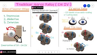
Trochlear Nerve Palsy | CN IV | Why Head T...
Med Madness
13,984 views

19:25
Path of the vagus nerve (anatomy)
Sam Webster
186,055 views

15:10
RS Ocular Motor Cranial Nerves Fourth Nerve
Michigan Medicine
9,983 views
24:57
Autonomic Nervous System (ANS) - Quick Rev...
Medicosis Perfectionalis
29,907 views
30:55
Optic Nerve & Visual Pathway | Cranial Ner...
Medicosis Perfectionalis
23,295 views

33:28
Neurology | Glossopharyngeal Nerve: Crania...
Ninja Nerd
172,130 views
11:39
Hypoglossal Nerve - The 12th Cranial Nerve...
Medicosis Perfectionalis
3,624 views

1:22:31
Facial Nerve | Cranial Nerve VII | Neurol...
Dr. Najeeb Lectures
154,312 views