Northern Blot Method - Animated Video
60.32k views1037 WordsCopy TextShare

Biology with Animations
I make animations in biology with PowerPoint, and this animation video is about a northern blot meth...
Video Transcript:
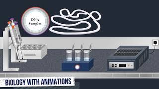
northern blood is a classic technique in molecular biology that reveals information about rna identity size and abundance and it can be used to study gene expression by detection of isolated mrna in a sample the first step in a northern blot is the preparation of the rna samples during this step formaldehyde is added to the samples for the denaturation of the rna molecules rna typically is a single stranded nucleic acid however the presence of self-complementary sequences in the rna strand leads to intrachain base pairing and folding of the ribonucleotide chain into complex structural forms known as
secondary structures formaldehyde contains a carbonyl group that reacts to form shift bases with the amino or amino groups of nucleobases these covalent adducts prevent normal base pairing and maintain the rna in a denatured state after preparation of the samples the rnas are separated by gel electrophoresis for the separation a loading buffer is added to the samples and used as a tracking dye which migrates in the same direction as rna allowing the user to monitor the progress of the separation agaris formaldehyde gel electrophoresis is most commonly used to separate mixtures of rna molecules during electrophoresis a
molecular weight size marker known as an rna ladder is commonly used to determine the size of rna molecules in the samples the rna ladder is added into well at one end of the gel once the molecular weight size marker is added the rna samples are loaded into wells then an electric current is applied to pull the samples through the gel based on their charge and size the rna molecules will travel through the gel at different speeds allowing them to be separated from one another the phosphate backbone of the rna molecule is negatively charged therefore when
placed in an electric field rnas will migrate to the positively charged anode because all rna molecules have the same amount of charge per mass small molecules move through the gel faster than large ones because the rna formaldehyde adducts are unstable formaldehyde must be present in the agarose gel to maintain the rna in the denatured state after the electrophoresis is complete the rna molecules in the gel can be stained when the gel is stained with an intercalating dye such as ethidium bromide the rna molecules can be seen under uv light as bands each representing a group
of same-sized rna molecules after electrophoresis the next step is northern blotting to transfer the rna molecules to the membrane a transfer buffer a solid support and a sheet of blotting paper acts as a wick for the transfer solution are used the wick is placed over the solid support in the transfer reservoir so the ends will be in the transfer buffer then it is wetted with the transfer solution next pieces of extra thick blotting paper are placed on top of the wick then they are wetted with the transfer solution next the gel is placed on the
thoroughly wetted wicking paper then a sheet of nylon membrane with the same size as the gel is pre-wetted with the transfer solution and placed on top of the gel next pre-wetted pieces of extra thick blotting paper are placed on top of the membrane the exposed areas of the wick are covered with strips of plastic wrap to prevent transfer buffer from bypassing the gel during the transfer process finally a dry stack of paper towels is placed on top of the membrane and gel then a glass plate is placed on top of these sheets paper with a
weight to maintain tight contact between the gel and membrane buffer transfer by capillary action from a region of high water potential to a region of low water potential is then used to move the rna from the gel onto the membrane consequently ion exchange interactions bind the rna to the membrane due to the negative charge of the rna and positive charge of the membrane the transfer is allowed to proceed overnight then once it is completed the blotting material and membrane are carefully removed from the gel next the membrane is briefly rinsed to remove any agaris that
may be stuck during the transfer then it is exposed to ultraviolet radiation to permanently attach the transferred rna to the membrane after attachment of the rna molecules to the membrane hybridization with radio-labeled probes is performed the membrane is placed in a bottle containing a pre-hybridization solution which is used to reduce non-specific hybridization with the probe next the bottle is incubated in hybridization oven at 42 degrees celsius for two hours once the incubation is complete the prehybridization solution is removed then a hybridization buffer is added into the bottle next labeled probes are added to the hybridization
solution commonly see dna is created with labeled primers for the rna sequence of interest to act as the probe which can be radioactively or fluorescently labeled once the hybridization probes are added the bottle is incubated overnight in the hybridization oven at 42 degrees celsius dna contains a large quantity of phosphorus in the fasfotiester linkages between nucleotides in the oligonucleotide chain dna can therefore be tracked by replacing its non-radioactive phosphorus with radioactive phosphorus 32. the radioactively labeled dna probes hybridized to their complementary sequences in the rna molecules after the hybridization of each probe to its target
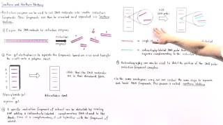
sequence the hybridization solution is removed next a wash buffer is added into the bottle then the membrane is incubated at 52 degrees celsius for 30 minutes the washing process is repeated three times to remove unbound and weakly binding probe after hybridization an autoradiography method is carried out to identify the location of radioactively labeled rna in the membrane the southern blot filter is placed inside a light proof cassette box then an x-ray film is laid over the top the cassette is closed and left for several hours to several days the radio isotope labeled rna exposes the
film which when developed shows a pattern of black bands that indicate the positions of labeled rna in the blot membrane subsequently this identification can be used to determine which rna molecule is present in each sample
Related Videos

11:46
Western Blot Method - Animated Video
Biology with Animations
122,969 views
10:01
Southern Blot Method - Animated Video
Biology with Animations
203,698 views
4:22
DNA Microarray Methodology
BioNetwork
292,532 views

14:40
Northern Blotting - Biology Tutorial
Joao's Lab
117,702 views

5:11
Northern Blotting
Frank Lectures
137,581 views

7:31
What is Gel Electrophoresis? | miniPCR bio™
miniPCR bio
372,884 views

12:40
Western Blot Protocol
SENS Research Foundation
52,434 views
3:47
Gas Chromatography - Flame Ionization Dete...
Biology with Animations
212,790 views

7:01
Northern Blot
Abnova
55,739 views

6:26
Southern Blot
Abnova
174,890 views

5:27
Agarose Gel Electrophoresis - Animated Video
Biology with Animations
127,616 views

3:56
PCR - Polymerase Chain Reaction (IQOG-CSIC)
CanalDivulgación
4,389,122 views

8:38
SDS-PAGE, Sodium Dodecyl Sulfate–PolyAcryl...
Biology with Animations
541,340 views
9:03
Southern and Northern Blotting
Andrey K
248,436 views

7:55
Gel Electrophoresis
Amoeba Sisters
2,239,950 views

9:11
Indirect ELISA Test - Animated Video
Biology with Animations
26,067 views

8:08
Southern Blotting
Frank Lectures
198,237 views

4:23
Western Blot / Protein Immunoblot explained
Henrik's Lab
315,736 views

18:00
Western blotting technique | principle and...
Shomu's Biology
444,577 views

8:02
Competitive ELISA Test - Animated Video
Biology with Animations
17,153 views