Anatomy of the Heart (Layers, Conducting System & Topography)
202.1k views2009 WordsCopy TextShare
Taim Talks Med
Content:
0:00 Introduction
0:29 Layers of the Heart
1:12 Endocardium
2:04 Myocardium
5:18 Epicardium...
Video Transcript:
What's up. Meditay here. Let's talk about the heart again.
In the last video, we covered the circulation system and the general anatomy of the heart. Now In this video, We're going to cover the Layers of the heart, which include the Endocardium, Myocardium, and Epicardium. Then we'll go through the conducting system.
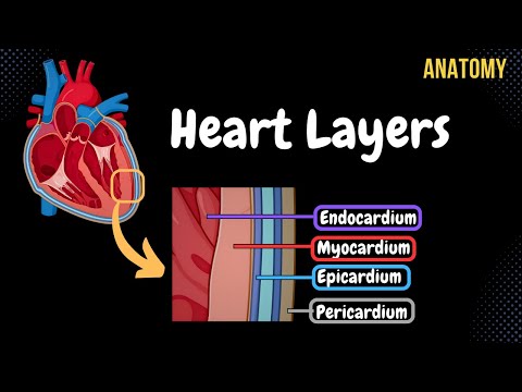
And after that, we're going to look at the general topography of the heart, which will help you from a clinical perspective. Let’s go through all of these starting with the layers of the heart. So here you see a raw picture of the heart without any type of coverings.
In real life, the heart is covered by a wet surface called serous pericardium. And then by another layer of dense connective tissue called fibrous pericardium. We’ll now start by cut the heart like this, so see all the layers.
Then we’ll take a small segment, and zoom in. Now we’re able to see all the layers of the heart, which include the endocardium, myocardium and the Epicardium. These three layers are what is considered a part of the actual heart.
The Serous and Fibrous pericardium we saw earlier are just coverings, meaning they’re not a part of the heart they’re just coverings. We’ll now go through all of these in a little more detail, starting with the endocardium. The endocardium is lined by endothelium just like the blood vessels, and it’s the innermost layer of the heart which means that it’s in direct contact with the blood.
One thing I want you to know about the endocardium is that it’s going to line the inner layer of the heart, but once it gets to the valves, it forms this double layer of endocardium, which I tried to show you here on the bicuspid valve. And this double layer of endocardium is the actual cusps you see in all the valves of the heart. It forms the cusps of the Bicuspid Valve, Tricuspid Valve, Pulmonary Valve and the Aortic Valve.
As well as the other valves we have like the valve of the inferior vena cava which is an embryonic valve, and the valve for the coronary sinus. So again what are the cusps of all of these valves made up of? It’s made up of a double layer of endocardium.
Cool. Underneath the endocardium, we’ll find the Myocardium. Myocardium consists of mainly muscles or muscle fibers, but you will also find a certain amount of connective tissue as well.
The first thing we need to understand is that the Myocardium will form the fibrous rings around each of the valves. It'll form the Right and Left Fibrous rings around the tricuspid valve and the mitral valve, and it'll form the fibrous rings around the pulmonary and the Aortic Valve, as you see here. This means that above the right and left fibrous rings, that's where the atria are.
And below the fibrous rings, that's where the ventricles are. And the reason why I’m telling you this is because the myocardium is arranged differently in the atria and the ventricles. So let’s do the myocardium in the atria first.
The Myocardium in the atria has two layers. The Superficial layer is a circular layer. And this circular layer is continuous.
So the way these muscle fibers are arranged is that they wrap around both atria, as you see here, and it kind of unites both atria from the outside. While the inner layer consists of longitudinal muscle fibers, and this one is individual for each atria. The Longitudinal muscle fibers form the inner muscle you'll see from the inside of the atrial wall, called Pectinate Muscle, remember this one?
So the Longitudinal muscle fibers form this muscle as well. So that's the Myocardium of the atria. Now let's do the Myocardium at the ventricles.
So the Myocardium at the Atria consists of two layers, right? The myocardium in the Ventricles, consists of 3 muscle fibers. We have the superficial longitudinal muscle layer, we have the deep longitudinal muscle layer.
And between those, there’s a middle circular muscle fibers. Let’s now schematically got through how these muscle fibers are distributed. So the superficial and the Deep longitudinal muscle fibers are connected.
And they're connected like this. so imagine on the left ventricle you have longitudinal superficial muscle fibers, right? It goes down to the Apex the Heart, it forms this vortex at the tip of the heart here.
Then it continue into the right ventricle as deep muscle fibers. So when I said that the deep and the superficial muscle fibers are connected, this is what I meant. It starts superficially and then forms a vortex at the apex and then goes deep at the other side.
Now same on the other side, it starts deeply and then goes to the Apex of the Heart and forms the vortex and then continues as superficial longitudinal fibers on the right side. This vortex it forms, this vortex at the Apex of the Heart, is called the Vortex Cordis, or vortex of the heart. The middle layer, or the circular layer, surrounds each ventricle as a see here.
And you'll notice that the left ventricle is much thicker than the right ventricle because the left ventricle has to pump the blood to the whole body, so it has to have more muscle in order to provide a more powerful contraction to supply the whole body. Here is a better animated picture of how the muscle fibers go. And really notice how the left ventricle is much thicker.
Now, remember the trabeculae carneae and papillary muscles inside the ventricles? The inner muscle layers for in the ventricle will form these muscles right here. So that's mostly it for the Myocardium.
The third layer of the heart is called the Epicardium. The epicardium is a little special because it's not a part of the heart itself, but since it sits so tight to the Myocardium, it's considered a part of the hearts layers. The epicardium is actually the visceral lamina of the serous pericardium, which is the covering of the heart.
So if this is the visceral lamina, or the epicardium. Here is the Myocardium, which the epicardium sits tightly onto. And the visceral lamina is connected to the other lamina of the serous pericardium up here, which is the parietal lamina.
So the Visceral lamina and the parietal lamina are separated across the whole heart, but they connect at the backside of the heart, where the great vessels are. So now, let's talk about the pericardium in a little more detail. So here we have a raw picture of a heart again.
But in real life, it has a layer called the Serous Pericardium. And the serous pericardium consists of two lamina or two plates. We have the visceral lamina of the serous pericardium, which you know is the epicardium closest to the heart, and the parietal lamina of serous pericardium directed towards the next pericardial layer, which is the Dense Fibrous Pericardium as you see here, which surrounds the heart and protects it.
Basically, the more superficially you get on the pericardium, the more fibrous the layers will be. Now between the visceral lamina and parietal lamina of the heart, there's a cavity. And this cavity is called Pericardial Cavity, which contains serous fluid.
This is a fluid that decreases the friction during the heart contraction. So it’s really crucial to have this serous fluid around the heart. But in real life, this cavity forms two pouches.
So if you turn the heart around like this. the first pouch it form is is Transverse Pericardial Sinus which lies between the ascending aorta and the pulmonary artery as you see here. The second pouch is the Oblique Pericardial Sinus, located on the heart's posterior aspect between the vena cava inferior and the Pulmonary Veins.
So that's mostly it for the walls of the heart. Now, let's do the quick anatomy of the conducting system. Now the heart's contraction happens within the heart itself, and the heart contains a particular type of muscle tissue that actually conducts the electrical impulses, and those electrical impulses come from a system of nodes and bundles inside the heart itself.
Now keep in mind that these structures are not anatomical. They're physiological, which means that you can't really physically see them with the eye, but they're within the heart. First, you have Sinoatrial node, located between the Right Auricle and the Superior Vena Cava up here.
The SA node generates impulses, and those impulses are generated to both atria and to the next node, the Atrioventricular Node, which is situated in the atrial septum between the ventricles and the atria. Then impulses are conducted through the Bundle of HIS, which divides into the right bundle branch and the left bundle branch. Which then conducts the impulse further as they divide and divide, where they now become the Purkinje fibers.
Which stimulates the muscle contraction. . So that fairly everything I had about the anatomy of the heart.
Let's now cover the Topography of the Heart. And we’ll do that by first going through the Holotopy of the Heart, meaning its location in relation to the body as a whole. So here you see the anterior view of the thorax.
The heart lies in the middle mediastinum. So if we cut the thorax in the middle and look at it in this direction, you'll see the middle Mediastium here in yellow, and that's where the heart is. So that's mainly the holotopy.
Next, we have the sceletopy, meaning the position of the heart in relation to the skeleton. So for that, let's use this picture. The upper border of the heart lies along the horizontal line of the level of the third rib, while the right border lies parallel to the sternal Margin Called the parasternal line, about (1.
5 cm) from the right side of the sternum. It goes from the 3rd rib to the 5th rib, and then the lower border goes obliquely like this. It goes from the cartilage of the 5th rib on the right side, to the apex of the heart, which lies in the level of the 5th intercostals space.
The left border of it runs from the apex cordis in the 5th intercostal space to the level of the 3rd rib. So now by looking at the heart's sceletopy, it's also important to know at which level the valves are, for examination purpose. So the atriventricular openings, that's the tricuspid and bicuspid valves, go from the sternal Junction of the 3rd rib on the left side to the sternal Junction of the 6th rib on the right side.
While the Aortic and the pulmonary openings go from the third sternal Junction on the left to the 4th sternal Junction on the right side. Alright, so, if you want to examine the status of each valve. Usually, you can hear them by using a Stethoscope and listen in the second intercostal space on the right side for the Aortic valve, and on the left side for the Pulmonary Valve.
While if you want to listen to the tricuspid and bicuspid valves, you can usually listen at the level of the 5th intercostal space. And here’s a picture that kinda sums up what we just went through. So that was mainly the sceletopy.
The last thing I want to talk about is the heart's syntopy, meaning the heart's position in relation to other organs. Anteriorly, it lies right behind the sternum, and posteriorly, you'll find the the oesophagus, and a lot fo vasculature. You know different types of blood vessels, nerves, and lymph tissue.
Laterally, you'll find theprotective layer of the lungs, called pleura. Then inferiorly, we have a diaphragm, and then superiorly, you have the great blood vessel, which are the Aorta and the Vena cava Superior. So that was mainly what I had about the anatomy of the heart in my last two videos, and I hope that was helpful.
Related Videos

21:55
Cardiovascular | Structures and Layers of ...
Ninja Nerd
1,189,039 views

48:01
Cardiovascular | Electrophysiology | Intri...
Ninja Nerd
1,651,766 views

28:32
The Cardiovascular System: An Overview
Strong Medicine
799,692 views

18:19
Anatomy of the Heart - External & Internal...
Taim Talks Med
643,149 views

1:12:50
Embryology | Development of the Heart ❤️
Ninja Nerd
1,227,540 views

18:32
Cardiovascular | Anatomy of the Heart | He...
Ninja Nerd
2,332,294 views

7:19
Heart Layers Anatomy Nursing: Pericardium,...
RegisteredNurseRN
44,503 views

4:23
The Difference Between Cardiac Arrest, Hea...
Health Decide
1,770,700 views
21:33
Cardiovascular System 1, Heart, Structure ...
Dr. John Campbell
6,426,039 views

29:46
ECG Interpretation Made Easy | ECG EKG Int...
ICU Advantage
476,509 views

44:59
Cardiovascular | Cardiac Output
Ninja Nerd
1,356,004 views

4:39
Heart Bypass Surgery (CABG)
Nucleus Medical Media
1,576,371 views

17:46
Heart Conduction System & ECG (EKG)
Siebert Science
180,245 views

9:52
Cardiovascular System 2, Heart, Blood flow...
Dr. John Campbell
983,163 views

23:59
Cardiovascular | Cardiac Cycle
Ninja Nerd
1,722,734 views

11:17
Circulatory System | Coronary Circulation
Ninja Nerd
605,112 views
6:08
Blood Flow Through the Heart (Made Easy in...
ICU Advantage
1,571,430 views

8:43
The Cardiac Cycle is SO EASY! Stop Making ...
Interactive Biology
1,326,920 views

26:26
Overview of Heart Anatomy Tutorial
The Noted Anatomist
392,804 views
3:54
Coronary Angioplasty (Femoral Access)
Nucleus Medical Media
26,922,596 views