Internal Spinal Cord (Gray Matter, White Matter, Funiculus) - Anatomy
181.84k views3556 WordsCopy TextShare
Taim Talks Med
PS! There's a little mistake in the table at the end (white matter). Numbers 1 and 2 are switched. A...
Video Transcript:
What’s up. Meditay Here. Let’s talk about the anatomy of the Central Nervous System.
In this segment, we will be talking about the Internal surface of the Spinal Cord. Basically go through everything you need to know regarding the anatomy of the tracts and nuclei within the spinal Cord. Alright, so the Central Nervous System consists of two parts.
The encephalon and the spinal Cord So in this is video, we’re first going to go through the internal surface of the Spinal Cord and talk about the distribution of white and grey matter within it. Then we’ll look detailed into the anatomy of the grey matter and the anatomy of the white matter. Then at the end of this video, I’ve made a quiz which you’ll hopefully be able to pass based on this video.
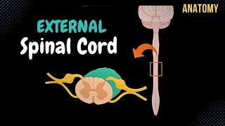
In our previous video, we looked at the Topography and the external structures of the Spinal Cord, as well as the anatomy of the spinal nerve and the two types of reflex arches we have through the spinal Cord. So if this is the first time you’re studying the spinal Cord, I recommend you to watch that first because this video will make much more sense with you knowing the external surface of the Spinal Cord. Alright.
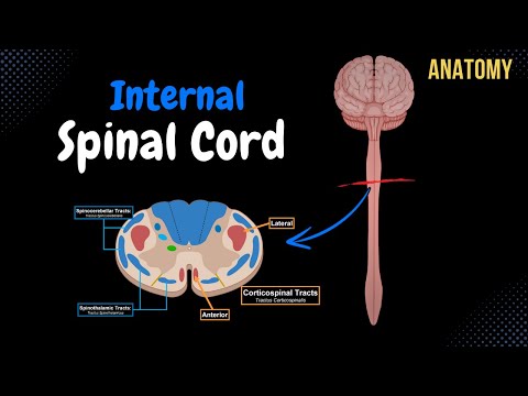
We can start by taking a small segment of the spinal Cord, and look at its internal surface. It’ll look like this. The internal surface of the Spinal Cord consists of grey matter and white matter.
The reason why they’re called grey and white matter is because if you look at a neuron. A neuron is colorless in general. And we know that because if you cut the spinal Cord physically and look at it underneath the microscope without any significant staining, you’ll see that the neurons are grey.
So the grey matter consists of nerve tissue rich in nerve cell bodies and dendrites. White matter consists of nerve tissue rich in myelinated axons and glial cells, as you see here. And the reason why myelinated axons are white is because they’re rich in lipid, and lipids are white.
And keep this in mind throughout this video because you’ll notice as we go through the structures in the grey matter, we’ll be talking about nuclei or nucleus because cell bodies are in the grey matter. When we’re talking about the white matter, we have structures called tracts, which are axons. So let’s start with the grey matter first.
The grey matter consists of three horns. We have an anterior horn and a posterior horn, or conu anterius and conu posterius. And where is the last horn?
The last horn is located in the spinal segments associated with the sympathetic and the parasympathetic fibers, between the C8 and L2, and S2 and S4 spinal cord segments. So if you cut the spinal Cord within those areas, you’ll see that we have an anterior horn, a posterior horn, and a lateral horn that give off either sympathetic fibers or parasympathetic fibers. So this segment is from an area outside of the sympathetic or parasympathetic segments.
-- Between the anterior and posterior horn, there’s the Intermediate column. Then in the middle, there’s the Central Zone, with the central canal in the middle. The central canal is filled with cerebrospinal fluid, which is the same fluid as in the subarachnoid space.
Around the central canal, there are cells called ependymal cells, forming a central gelatinous substance of the Spinal Cord, which is the same lining as the rest of the ventricles in the spinal Cord. So now I wanna focus on each of these zones, basically go through the most important nuclei you’ll find, and we’ll start with the Anterior horn. So the anterior horn is pretty easy as it primarily consists of motor nuclei.
Their axons leave the spinal Cord as the motor root of the spinal nerve. So that’s mostly it for the anterior horn. The posterior horn is associated with receiving sensory information.
You’ll notice that it consists of several nuclei responsible for certain types of sensory information. So the first one is the marginal nucleus consisting of interneurons triggered by any sensory neuron coming into the spinal Cord. Then there’s the gelatinous substance, which also consists of interneurons that modulate sensory input Then there’s the Nucleus Proprius, which consists of neurons that modulate sensory input like pain, touch, and temperature.
They take the information and send them upwards to the higher senses After that, we have the posterior thoracic nucleus, which receives unconscious proprioceptive movement. Basically giving information about the position and posture of the body. So that was the posterior horn.
Now let’s do the intermediate zone. And the first nucleus here is the intermediomedial nucleus since it’s located medially within the grey matter. This nucleus has more or less the same function as the posterior thoracic nucleus, which is unconscious proprioception.
And some sources consider the posterior thoracic nucleus as a part of the intermediate zone, not a part of the posterior horn, so keep that in mind. But there are two more nuclei within the intermediate zone, and they’re located within the lateral horn. Remember we mentioned this earlier?
So in the lateral horn, we’ll find two different nuclei depending on where we are within the spinal Cord. If we’re between spinal segments C8 through L2, then we have the IntermedioLateral nucleus, which consists of sympathetic fibers for the fight or flight response. They will send their fibers together with the motor fibers down through the anterior root of the spinal nerve.
If we’re looking at a segment between S2 and S4, we will see the Sacral Parasympathetic nuclei, which also send their fibers through the anterior root of the spinal nerve, responsible for the rest and digest state of the person. So the intermediate column consists of the Intermediomedial nucleus, Intermediolateral nucleus, Sacral parasympathetic nucleus, and the posterior thoracic nucleus if your sources say so. Alright, so now we’ve covered the majority of nuclei within all three zones of the grey matter.
Along the outer part of the grey matter, you’ll find a very thin layer of white matter called fasciculi proprii, which are fibers that connect adjacent parts of the grey matter together or adjacent segments because you can also divide the grey matter into segments. Another thing you’ll find is grey matter that is pressed into the white matter called spinal reticular formation, or Formatio retucularis spinalis, which continues upwards into the brainstem. It consists of neurons that make a communicative network for activating certain sensory information for basic living.
So it regulates respiration, heartbeat, blood pressure, and all of those things. Here’s maybe a better representation of the spinal reticular formation, and again it goes upwards into the brainstem. So that was all for the grey matter of the Spinal Cord, now let’s do the white matter.
The white matter of the Spinal Cord, remember its nerve tissue rich in myelinated axons, so we’re not talking about nucleus anymore. We’re talking about tracts or bundles of fibers. And the white matter is divided into three portions because you’ll find connective tissue separating these three portions.
We have the Posterior Funiculus, the Lateral funiculus and the Anterior funiculus. So again, these funiculi are bundles of fibers that are surrounded by connective tissue. Now before we go detailed into each of these funiculi.
I wanna spend a little bit of time explaining the general arrangement of fibers within the white matter. And by understanding that, the tracts will get much more logical. Alright, so nerve fibers are arranged in bundles or tracts, right?
So here are many neurons. There are connective tissue around all of those neurons, and that was what we call a tract. And there are three .
. um. .
directions these tracts can go. They can either go upwards, as ascending tracts, or afferent tracts, remember a stands for arrive, so these tracts arrive to the brain, but they can also descend, go down as descending tracts, or efferent tracts e stands for exit. So the asceding tracts receive sensory information from anywhere in the body, and send them up to your higher senses to make sense out of them.
And then once you’ve done that, you’d wanna react to the sensory information, or you just wanna move a muscle in general, so you activate the descending tracts. So you accidentally put your hand above something hot, sensory information is sent up, and then you react by descending tracts activating muscles to remove your hand. And again, all of that happens through funiculi, which means long ropes, found within the posterior, lateral, and anterior parts of the Spinal Cord.
The ascending and descending tracts can further be divided into two parts. The ascending tracts are generally divided into unconscious and conscious sensory information. Now, what is the difference?
Well, everything that goes to the cortex of your cerebrum is considered conscious. Unconscious sensory input goes to the cerebellum. The cerebellum is an organ responsible for balance and posture, so the unconscious ascending tract relays unconscious proprioceptive sensation.
You’ll see this word a lot as you study the tracts of the central nervous system. So it’s important that you have a general knowledge about it. Unconscious proprioception is responsible for posture, meaning sensory fibers from muscles are all the time sent to the cerebellum so that it can activating the necessary muscles to keep your posture, as well as activating muscles that support your joints and the natural position of your limbs, so information about joint stability is considered unconscious proprioception as well.
But there is one more thing. Imagine you’re outside, walking and minding your own business. All of a sudden, YOU SEE A CAR DRIVING TOWARDS YOU AND BREAKS RIGHT IN FRONT OF YOU.
Your initial response is tensing your muscles, a process called feedforward control. Your muscles tenses due to something in the external environment. And in the majority of times, you’re not in control of it, and we consider this as unconscious proprioception as well.
They’re unconscious because they go to the cerebellum, which is this organ right here. In our spinal Cord, we have tracts called the anterior spinocerebellar tract and the lateral spinocerebellar tract. They end with the word cerebellar, so they go to the cerebellum.
Conscious sensory information relays conscious proprioception, like kinesia, which is conscious muscle movement. The conscious joint position is considered a part of this system, as well as a sense of force. Meaning as your standing, you consciously know that there’s a force acting against you, which is the floor against your feet.
The conscious sensory information is also responsible for sensing certain things, like touch, and pain and pressure and temperature. Examples of conscious ascending tracts are the anterior and lateral spinothalamic tracts, the cuneate, and the gracilis fascicle, which send their information to the cortex of the cerebrum. So that is the ascending tracts, but we also divide the descending tracts into two parts.
We divide it into Involuntary movements and voluntary movements. So one of the important parts of our cerebral cortex is the primary motor cortex. If we look at it underneath the microscope, you’ll find that it consists of pyramidal cells.
They’re pyramidal because they literally look like pyramids. So when you decided you wanted to click on this video or search for the internal surface of the Spinal Cord on youtube, you activated the pyramidal cells to give the command to your muscles. All the tracts that come from the pyramidal cells are called voluntary movements.
And they help you make fine conscious movements like when you’re writing with a pen. These movements are so precise that they make your handwriting look good. These tracts usually start with the name Cortico- Like the anterior corticospinal tract and the lateral corticospinal tract.
When motor fibers doesn’t come from the pyramidal cells, they’re called extrapyramidal tracts meaning these tracts originate from other parts of your brain instead of the primary motor cortex So these movements are movements you don’t really think about, like keeping your balance and posture when you’re walking, as well as rough movements or coarse movements. These tracts doesn’t come from the primary motor cortex, so they have other names like rubrospinal tract, tectospinal tract and olivospinal tracts. So I hope this made a little sense to you all because once you’ve visualized this part.
The next part of this video will be much easier to understand. So let’s now start by talking about the tracts within each of these funiculi, and we’ll start with the posterior one. Ok, so in this diagram, I’ve made the blue colors represent sensory fibers and the red ones representing motor fibers.
The posterior funiculus has two sensory tracts. The first one is called Fasciculus Gracilis. This tract will conduct impulses from the lower part of the trunk and the lower limbs, so it’s present in all the segments of the Spinal Cord, and it takes the information to the cortex.
And since it brings info to the cortex, then remember its function is conscious sensation. In this case, it’s responsible for the epicritic sensibility of the lower parts of the body. Epicritic sensibility means Conscious proprioception, which remember is Kinesia, joint position, and sense of force.
But epicritic sensibility also means receiving info from the mechanoreceptors. Now, what does that mean? It senses two-point discrimination, so the minimal distance between two touchpoints, until you actually sense that there are two objects touching you, that is the two points discrimination.
Mechanoreceptors also sense vibration and touch. So all of those things are what we call epicritic sensibility. The other ascending tract of the posterior funiculus is the Fasciculus Cuneatus, which brings sensory information from the upper body and sends it to the cortex.
So it does exactly the same as Fasciculus Gracilis, just for the upper body. And since this tract si only for the upper body, you’ll find this tract only above the thoracic spinal segment number 6. So that was all of the posterior funiculus.
Fasciculus Gracilis for lower limb and Fasciclus Cuneatus for Upper limb. I use the letter G in Gracilis as Genitals to remember that gracilis is for the lower part of the body Next, Let’s do the Lateral and the anterior funiculus together because you’ll find tracts that do the same but are present on both the lateral and the anterior funiculus. Just remember that they’re divided by connective tissue.
These two funiculi are divided by connective tissue, and we’ll represent the connective tissue using these two brown lines. The first tracts are ascending tracts located on the lateral funiculus, called the SpinoCerebellar tracts. We have two, there’s the anterior spinocerebellar tract and the posterior spinocerebellar tract.
Before ascending up, the posterior spinocerebellar tract receives sensory input from the posterior thoracic nucleus, and the anterior spinocerebellar tract receives sensory input from the intermediomedial nucleus. They then ascend to the cerebellum because they both end with the word cerebellum. And remember, if something ascends to the cerebellum and not the cerebral cortex.
Then they provide unconscious proprioception, which later will provide unconscious contraction of muscles for posture, joint stability, and the feedforward control system. So that is the spinocerebellar tract. Next, we have the spinothalamic tracts, and there are two spinothalamic tracts in our spinal Cord.
There’s the anterior spinothalamic tract in the anterior funiculus and a lateral spinothalamic tract in the lateral funiculus. These two tracts are formed by axons coming from the nucleus proprius, and then they ascend to the cortex. And since they go to the cortex, they are responsible for the conscious sensation of pain, temperature, pressure, and touch.
So that was all the sensory tracts of the Spinal Cord. Now let’s do the motor tracts or the descending tracts. This includes the corticospinal tracts.
We have two corticospinal tracts. There’s the Anterior corticospinal tract in the anterior funiculus and the lateral corticospinal tracts in the lateral funiculus These tracts start with the name ‘’Cortico’’, meaning they come from the pyramidal cells of the primary motor cortex. So they are pyramidal tracts.
Now the Lateral and Anterior corticospinal tracts descend differently, but they both synapse with the motor nuclei to stimulate the motor fibers in the anterior root of the spinal nerve. Alright. So the Lateral Corticospinal tract starts off at the cortex and then descend, but they decussate at the medulla oblongata, meaning they cross to the other side as you see here at the medulla oblongata, and form the decussation of pyramids, and then they descend further as the lateral corticospinal tract in the spinal Cord and synapse with the motor nuclei.
The Anterior corticospinal tract is a little bit different in that they descend without crossing. Instead, they cross at each segment they’re going to leave from. So in this example, if they’re gonna leave at this segment, they cross to the other side and synapse with the motor nuclei.
And since they come from the cortex, they’re responsible for conscious movement. So that’s the corticospinal tracts Next, we have the Rubrospinal tract. Rubro means red, and the reason why they’re called rubrospinal tract Is because we have red nuclei located inside the midbrain of the brainstem.
So these fibers are extrapyramidal fibers because they originate from the red nucleus of the midbrain and go down as rubrospinal tract. And remember, extrapyramidal tracts are responsible for fine coordination of movements and support voluntary movements. They make our voluntary movements more precise.
SO that is the rubrospinal tract. The next tract is the tectospinal tract, located in the anterior funiculus. It transmits motor impulses for the eyes and neck muscles meaning they coordinate the eyes and the neck muscles when you look at something.
I’ve animated this very badly, but imagine you’re looking at a hamburger, you look at it, and you keep looking at it as it passes you, and your neck muscles follow your eyes. That’s what this tectospinal tract is responsible for. It’s called tectospinal tract because it comes from the tectum of the midbrain, it’s located on the posterior surface of he midbrain, which is on your brainstem, and then descend down and synapse with the motor nuclei.
It’s extrapyramidal, so it unconsciously moved your neck muscles with your eyes. Next is the vestibulospinal tract. Inside of your ear, the inner ear, you have a system called the vestibular system.
The vestibular system has crystals within it sensing the position of your head, whether your head is tilted upside down or to the side, all of that is sensed and through nerves, it’s sent to the brainstem, and then down to your spinal Cord to keep your balance and posture. So the vestibulospinal tract is responsible for keeping your balance and posture. So if you’re running and trip on a rock and you’re about to fall, your vestibular system, along with other systems, will quickly react to that and quickly activate the necessary muscles in order for you to keep your balance.
And this happens involuntarily because this tract doesn’t come from your cortex. It comes from your brainstem. So that is the Vestibulospinal tract.
Next, we have the olivospinal tract. The olivospinal tract is also responsible for balance and posture. Inside of your medulla oblongata, there are olivary nuclei that send out fibers to the spinal Cord as the olivospinal tract, which also synapse with the motor nuclei.
Keep that in mind all motor tracts will synapse with the motor nuclei of the grey mater. Then we have the Reticulospinal Tract. We have a lateral Reticulospinal tract and a medial reticulospinal tract which are also a part fo the balance and posture system.
They come from the reticular formation inside the brainstem and also from the reticular formation of the Spinal Cord, remember we talked about that earlier? Just to remind you again. The Reticular system are responsible for Sleep, alertness, cardiovascular control, breathing and all of those vital things.
But they’re also responsible for motor control like your balance and posture, through the reticulospinal tract. So that is all of these. Then lastly there’s the medial longitudinal fascicle, located in the anterior fasciculus only in the cervical segments, which coordinates involuntary movements of the head neck and eyes through synapses between the cranial nerves.
And then they’re going to synapse with the motor nuclei of the grey matter as well. So that was all I had for the grey and the white matter of the spinal cord. So I made this table for the nuclei in the grey matter And this is where this video gets scary, I am going to make all of the names disappear.
Now can you, from the beginning, tell me what is the name of number 1, what is the name of number 2, and so on. And here is one for the white matter as well. You can pause the video if you want and have a look, and then here is an empty table for you to fill.
So that was everything I had for the anatomy of the Spinal Cord. If you found this video helpful, please put a like, comment, share, whatever you find convenient to you. The next video is going to be about the Medulla Oblongata.
Related Videos

16:46
Medulla Oblongata Anatomy - External & Int...
Taim Talks Med
265,103 views
15:57
External Spinal Cord (Surface, Segments, S...
Taim Talks Med
175,135 views
8:03
FSc Part 1 Mathematics Ex 3.5 Q # 1 Use C...
Dr Sajjad Khan Math Academy
7 views

17:50
Spinal Cord Pathways
Dirty Medicine
345,402 views

19:46
Trump on Upholding Constitution: "I Don't ...
The Daily Show
4,255,430 views

21:33
Internal Cerebrum (Association, Commissura...
Taim Talks Med
194,775 views
20:35
Brachial Plexus (Scheme + Quiz) | Anatomy
Taim Talks Med
77,798 views

2:28:58
Shaolin Warrior Master: Hidden Epidemic No...
The Diary Of A CEO
1,701,883 views

37:14
Neurology | Spinal Cord: Gray Matter Struc...
Ninja Nerd
387,738 views

16:57
Corticospinal tract
The Noted Anatomist
165,612 views
18:19
Anatomy of the Heart - External & Internal...
Taim Talks Med
673,438 views
22:57
Neurology | Spinal Cord Blood Supply
Ninja Nerd
144,029 views
35:26
Pain Physiology | Nociception
Dr Matt & Dr Mike
32,178 views
13:12
Action Potential in the Neuron
Harvard Extension School
3,119,342 views

20:12
What Symptom Does Each Multiple Sclerosis ...
Dr. Brandon Beaber
110,730 views

25:08
Introduction to the gastrointestinal tract...
Sam Webster
99,820 views

18:18
Cranial Nerve BASICS - The 12 cranial nerv...
ICU Advantage
1,680,375 views

35:16
Foundational features of the brainstem
The Noted Anatomist
394,170 views

3:14:02
Spinothalamic Tract | Ascending Tracts | S...
Dr. Najeeb Lectures
5,085,557 views

16:52
Pons - External and Internal (White & Grey...
Taim Talks Med
181,836 views