Cardiac Development by L. McCabe | OPENPediatrics
519.31k Aufrufe1064 WörterText kopierenTeilen

OPENPediatrics
This lecture presents important features of cardiac development.
Please visit: www.openpediatrics.o...
Video-Transkript:
Cardiac development, by Lisa McCabe. Hello, my name is Lisa McCabe. I’m a clinical nurse specialist at Children’s Hospital Boston in the cardiovascular program.
I will be discussing with you today Cardiac development. The information I swill share is consistent with our practice here at Children’s Hospital Boston. You may want to adapt this information to your own institutional practice.
Fetal Development. During the first week of fetal life, the fertilized egg develops into a blastocyte and implants in the mother’s uterus. During the second week of fetal life, the blastocyte implants deeper into the uterine wall, and a primitive placenta begins to form.
During the third week of fetal life, the primitive umbilical cord develops. Also at this time, the blastocyte develops into a three-layered disk. The three layers are: the endoderm, mesoderm and ectoderm.
Specific body systems will develop from each layer. The endoderm, or inner layer, gives rise to the primitive intestinal tube, mucous membranes, glands, lung buds, urinary tract, and yolk sac. The mesoderm, of middle layer, gives rise to the heart and vascular system, the dermis, subcutaneous tissue, muscles, skeleton, sex glands, lymph glands, kidneys, connective tissue, and blood cells.
And finally the ectoderm, or outer layer, gives rises to the epidermis, hair, sebaceous glands, sweat glands, and nervous system. Cardiogenesis. Early in the development, the primitive heart develops two tubes that merge into one tube.
The single tube begins to swell, and develops into various anatomic features of the heart. The heart begins o beat by week three. In normal cardiac development, the cardiac tube will twist and turns on itself in a rightward direction.
This is called dextral-looping. This results in the right ventricle developing on the right side of the heart and the left ventricle developing on the left side of the heart. Abnormal looping in a leftward direction is called leval-looping.
This results in the right ventricle developing on the left side of the heart and the left ventricle developing on the right side of the heart. Atrial Septation. Entering into the fifth week of fetal life, the atrial septum and ventricular septum begins to form.
The atrial septum grows in layers, and includes the tissues of septum premium, septum secondo, and endocardial cushion tissue. The endocardial cushion tissue is located in the middle of the heart. From this tissue arises the tricuspid valve, mitral valve, part of the atrial septum, and part of the ventricular septum.
Septum prium grows downward between the right and left atrium, and eventually fuses with the endocardial cushion tissues. The septum secudnum grows parallel to septum primum. Both septum primum and septum secundum develop with holes in them to create a passageway for blood to flow from right atrium to left atrium through the foramen ovale.
As long as the pressures on the right atrium are higher than pressures in the left atrium, the passage will stay open to allow the blood to flow from right to left. Errors may occur during atrial septation. A secundum atrial septal defect is an opening in the middle of the atrial septum, produce when the tissue of septum primum does not reach septum secundum adequately to completely close the wall or when a tiny hole is left in the septum primum as the tissues grow.
A primum atrial septal defect is an opening in the lower part of the atrial septum, near the tricuspid and mitral valves. It results from problems in the growth of the endocardial cushion tissues. A defect here is often associated with a defect in the mitral valve, as well as tricuspid valve.
The most severe form of this is an atrial ventricular canal defect. Truncal Septation. The aorta and pulmonary artery develop from a single tubular structure: the truncus arteriosis.
Two areas of thickened tissue project into the lumen of this tube on the right and left side. As they continue to grow in size, these ridges meet and fuse to form the septum, which takes a spiral course toward the end of this tube. This septum divides the truncus into two vessels: the aorta and the pulmonary artery.
The conal tissue involved in the septation of the truncus arteriosis also directs placement of the aorta and pulmonary artery over their related ventricles. Ventricular Septation. By the fifth week of fetal life, the atria completely separate into the right atrium and left atrium.
Also at this time, the ventricular septum continues to develop. The muscular portion of the ventricular septum develops from the apex of the common ventricle. The membranous septum develops from the endocardial cushion tissues.
Conal tissue contributes to the complete closure of the septum between the aortic and pulmonary valves. Errors may occur during ventricular septation. Muscular defects can occur in any portion of the muscular septum.
Small defects often close on their own as the tissues develop. Membranous defects are located behind the septal leaflet of the tricuspid valve. Sub-pulmonary defects are caused by deficiency of the conal septum.
Atrial ventricular canal type defects are caused by defects in the endocardial cushion tissues of the intraventricular septum. Arch Formation. The aorta and pulmonary artery and their branching vessels are formed from the brachial arch arteries.
Initially there are six pairs of brachial arch arteries. The first, second, and fifth pairs disappear, forming ligaments that hold the heart in place. The third aortic arch forms the common carotid artery, external carotid artery, and internal carotid artery.
The fourth aortic arch forms part of the final aortic arch, as well as the proximal portion of the right subclavian artery. The sixth aortic arch produces the proximal segment of the branch pulmonary arteries, the ductus arteriosis, and provides pulmonary blood flow via the branch that develops lung buds. Errors may occur in arch formation that result in the following defects: coarctation of the aorta, interrupted aortic arch, aortic atresia, patent ductus arteriosis, and vascular ring.
In just a few short weeks, the human heart has undergone major development. By the third week of fetal life, the heart is beating. BY day 44, the fetal heart resembles the postnatal heart in structure and function.
It will continue to grow and develop over the next 30 to 32 weeks. Many of the features that may results in a heart defect, however, have also begun to develop. Please help us improve the content by providing us with some feedback.
Ähnliche Videos

11:48
Development of the Heart | The Heart Tube ...
Byte Size Med
382,253 views
11:52
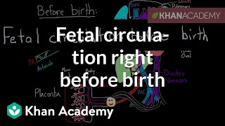
Fetal circulation right before birth | Cir...
khanacademymedicine
1,782,232 views

Healing Forest Ambience | 528Hz + 741Hz + ...
Healing Energy Frequency

8:43
The Cardiac Cycle is SO EASY! Stop Making ...
Interactive Biology
1,326,865 views

15:48
Chest Physiotherapy by S. Hamilton | OPENP...
OPENPediatrics
414,791 views

1:12:50
Embryology | Development of the Heart ❤️
Ninja Nerd
1,227,519 views

1:47:45
Gentle music, calms the nervous system and...
Soothing Soul
5,609,226 views
8:14
Circulatory System and Pathway of Blood Th...
Amoeba Sisters
5,023,456 views

29:54
3D Body Cavities Embryology Part 1: Embryo...
MedicoVisual - Visual Medical Lectures
79,644 views

19:10
Heart Murmurs and Heart Sounds: Visual Exp...
Zero To Finals
1,923,518 views

55:51
3D Heart Tube Embryology - Myoepicardial m...
MedicoVisual - Visual Medical Lectures
70,147 views

23:14
Anatomy of the heart
Osmosis from Elsevier
87,316 views

26:20
Gastrointestinal | Development & Embryolog...
Ninja Nerd
1,040,170 views

Tranquill Jazz In Lakeside | Living Coffee...
Tranquill Jazz Melody

1:02:52
Embryology | Development of Vascular System
Ninja Nerd
367,359 views

11:07
Foetal (Fetal) Circulation
Armando Hasudungan
1,610,958 views

47:48
Embryology for the Cardiologist (Regina La...
Houston Methodist DeBakey CV Education
7,467 views

15:33
Vasculogenesis and Angiogenesis - Blood Is...
MedicoVisual - Visual Medical Lectures
49,052 views
10:14
Development of the Respiratory System | St...
Byte Size Med
183,071 views
6:09
Embryology: from Fertilization to Gastrula...
Alila Medical Media
621,835 views